

Quantitative multi-modal MR imaging as a non-invasive prognostic tool for patients with recurrent low-grade glioma
- PMID: 28124178
- PMCID: PMC5373029
- DOI: 10.1007/s11060-016-2355-y
Quantitative multi-modal MR imaging as a non-invasive prognostic tool for patients with recurrent low-grade glioma
Abstract
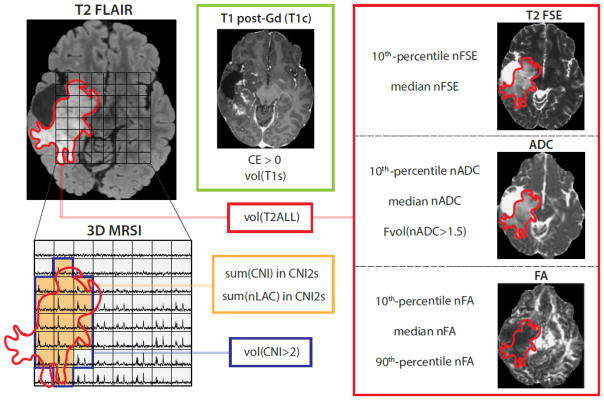
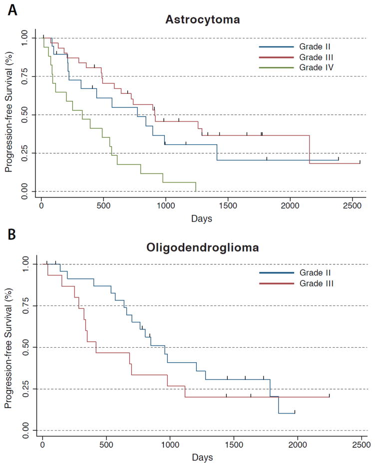
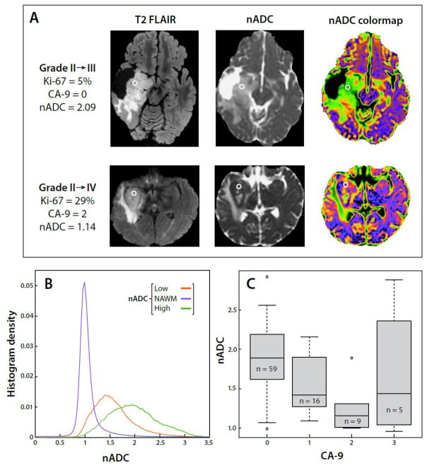
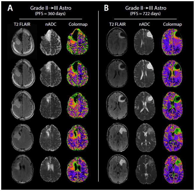
Low-grade gliomas can vary widely in disease course and therefore patient outcome. While current characterization relies on both histological and molecular analysis of tissue resected during surgery, there remains high variability within glioma subtypes in terms of response to treatment and outcome. In this study we hypothesized that parameters obtained from magnetic resonance data would be associated with progression-free survival for patients with recurrent low-grade glioma. The values considered were derived from the analysis of anatomic imaging, diffusion weighted imaging, and 1H magnetic resonance spectroscopic imaging data. Metrics obtained from diffusion and spectroscopic imaging presented strong prognostic capability within the entire population as well as when restricted to astrocytomas, but demonstrated more limited efficacy in the oligodendrogliomas. The results indicate that multi-parametric imaging data may be applied as a non-invasive means of assessing prognosis and may contribute to developing personalized treatment plans for patients with recurrent low-grade glioma.
Keywords: Diffusion; Glioma; MRI; Progression-free survival.
Figures
Similar articles
-
Prognostic Value of O-(2-[18F]-Fluoroethyl)-L-Tyrosine-Positron Emission Tomography Imaging for Histopathologic Characteristics and Progression-Free Survival in Patients with Low-Grade Glioma.World Neurosurg. 2016 May;89:230-9. doi: 10.1016/j.wneu.2016.01.085. Epub 2016 Mar 9. World Neurosurg. 2016. PMID: 26855307
-
Magnetic resonance analysis of malignant transformation in recurrent glioma.Neuro Oncol. 2016 Aug;18(8):1169-79. doi: 10.1093/neuonc/now008. Epub 2016 Feb 23. Neuro Oncol. 2016. PMID: 26911151 Free PMC article.
-
Optimizing a machine learning based glioma grading system using multi-parametric MRI histogram and texture features.Oncotarget. 2017 Jul 18;8(29):47816-47830. doi: 10.18632/oncotarget.18001. Oncotarget. 2017. PMID: 28599282 Free PMC article.
-
Advances in Magnetic Resonance Imaging and Positron Emission Tomography Imaging for Grading and Molecular Characterization of Glioma.Semin Radiat Oncol. 2015 Jul;25(3):164-71. doi: 10.1016/j.semradonc.2015.02.002. Epub 2015 Feb 23. Semin Radiat Oncol. 2015. PMID: 26050586 Review.
-
Susceptibility-Weighted Imaging of Glioma: Update on Current Imaging Status and Future Directions.J Neuroimaging. 2016 Jul;26(4):383-90. doi: 10.1111/jon.12360. Epub 2016 May 26. J Neuroimaging. 2016. PMID: 27227542 Review.
Cited by
-
Increasing FLAIR signal intensity in the postoperative cavity predicts progression in gross-total resected high-grade gliomas.J Neurooncol. 2018 May;137(3):631-638. doi: 10.1007/s11060-018-2758-z. Epub 2018 Mar 21. J Neurooncol. 2018. PMID: 29564748
-
Relationship of In Vivo MR Parameters to Histopathological and Molecular Characteristics of Newly Diagnosed, Nonenhancing Lower-Grade Gliomas.Transl Oncol. 2018 Aug;11(4):941-949. doi: 10.1016/j.tranon.2018.05.005. Epub 2018 Jun 18. Transl Oncol. 2018. PMID: 29883968 Free PMC article.
-
MR image phenotypes may add prognostic value to clinical features in IDH wild-type lower-grade gliomas.Eur Radiol. 2020 Jun;30(6):3035-3045. doi: 10.1007/s00330-020-06683-2. Epub 2020 Feb 14. Eur Radiol. 2020. PMID: 32060714
-
An automated approach for predicting glioma grade and survival of LGG patients using CNN and radiomics.Front Oncol. 2022 Aug 12;12:969907. doi: 10.3389/fonc.2022.969907. eCollection 2022. Front Oncol. 2022. PMID: 36033433 Free PMC article.
-
Automated Detection of Brain Tumor through Magnetic Resonance Images Using Convolutional Neural Network.Biomed Res Int. 2021 Nov 30;2021:3365043. doi: 10.1155/2021/3365043. eCollection 2021. Biomed Res Int. 2021. PMID: 34912889 Free PMC article.
References
-
- Grier JT, Batchelor T. Low-Grade Gliomas in Adults. The Oncologist. 2006;11(6):681–693. - PubMed
Publication types
MeSH terms
Grants and funding
LinkOut - more resources
Full Text Sources
Other Literature Sources
Medical
