. 2017 Apr;40(2):645-656.
doi: 10.1007/s10753-017-0511-y.

3'UTR AU-Rich Elements (AREs) and the RNA-Binding Protein Tristetraprolin (TTP) Are Not Required for the LPS-Mediated Destabilization of Phospholipase-Cβ-2 mRNA in Murine Macrophages

Affiliations

3'UTR AU-Rich Elements (AREs) and the RNA-Binding Protein Tristetraprolin (TTP) Are Not Required for the LPS-Mediated Destabilization of Phospholipase-Cβ-2 mRNA in Murine Macrophages

Smita Shukla et al. Inflammation. 2017 Apr.

Abstract

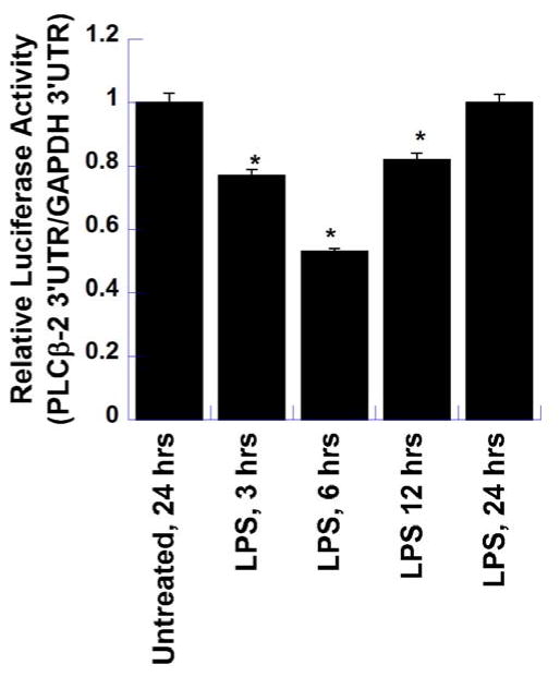
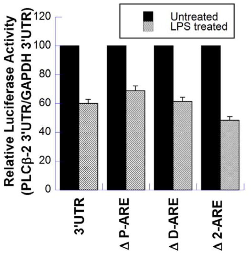
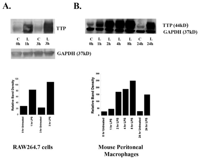
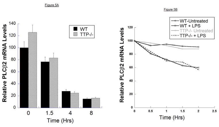
We have shown previously that bacterial lipopolysaccharide (LPS)-mediated suppression of phospholipase-Cβ-2 (PLCβ-2) expression is involved in M1 (inflammatory) to M2-like (wound healing) phenotypic switching of macrophages triggered by adenosine. This suppression is mediated post-transcriptionally by destabilization of PLCβ-2 mRNA (messenger ribonucleic acid). To investigate the mechanism of this LPS-mediated destabilization, we examined the roles of RNA-binding agents including microRNAs and RNA-binding proteins that are involved in regulating stability of mRNAs encoding growth factors, inflammatory mediators, and proto-oncogenes. Adenylate and uridylate (AU)-rich elements (AREs) in 3'UTRs are specific recognition sites for RNA-binding proteins including tristetraprolin (TTP), HuR, and AUF1 and for microRNAs that are involved in regulating mRNA stability. In this study, we investigated the role of TTP and AREs in regulating PLCβ-2 mRNA stability. The 3'UTR of the PLCβ-2 gene was inserted into the pLightswitch luciferase reporter plasmid and transfected into RAW264.7 cells. LPS suppressed luciferase expression from this reporter. Luciferase expression from mutant 3'UTR constructs lacking AREs was similarly downregulated, suggesting that these regions are not required for LPS-mediated suppression of PLCβ-2. TTP was rapidly upregulated in both primary murine macrophages and RAW264.7 cells in response to LPS. Suppression of PLCβ-2 by LPS was examined using macrophages from mice lacking TTP (TTP-/-). LPS suppressed PLCβ-2 expression to the same extent in wild type (WT) and TTP-/- macrophages. Also, the rate of decay of PLCβ-2 mRNA in LPS-treated macrophages following transcriptional blockade was similar in WT and TTP-/- macrophages, clearly indicating that TTP is not involved in LPS-mediated destabilization of PLCβ-2 mRNA in macrophages.

Keywords: 3’UTR; AU-rich elements; lipopolysaccharide (LPS); macrophages; phospholipase-Cβ-2; tristetraprolin.


Figures

Figure 1
Figure 1. Bioinformatic Analysis of PLCβ-2 3′UTR
A. The PLCβ-2 3′UTR (GenBank NM_177568.3) is 1481 bp long, and harbors two AU–rich elements (AREs) shown in bold. One is the canonical pentamer AUUUA, and the other has two overlapping pentamers. AREs are putative binding sites for RNA-binding proteins such as, TTP, HuR and AUF1 which are involved in regulation of mRNA stability. B. Analysis using TargetScan 7.1 shows that the second ARE is conserved across several mammalian species. Dashes indicate where sequences do not match.
Figure 2
Figure 2. PLCβ-2-3′UTR Regulation of Luciferase Reporter Gene Expression by LPS
To test the role of PLCβ-2 3′UTR in the destabilization of PLCβ-2 mRNA by LPS, a luciferase reporter assay was performed using two different plasmid constructs. The first contained the PLCβ-2 3′UTR downstream of the luciferase gene, and the other the GAPDH 3′UTR down-stream of the luciferase gene to serve as control. RAW264.7 cells were transfected with these constructs and treated with LPS for various time-periods. One set of wells was left untreated to provide constitutive luciferase expression. All samples were harvested at 24 hours and luciferase assays were performed using equal volumes of lysates and Renilla Glo. The results show the relative expression of luciferase from the PLCβ-2 3′UTR construct versus that of the GAPDH 3′UTR construct, and are the means ± S.D. of two separate experiments, each performed in triplicate. (* P_ <0.01 in comparison to Untreated Control)
Figure 3
Figure 3. Effect of Deletions of Proximal and Distal AREs in the PLCβ-2-3′UTR on the LPS-mediated suppression of luciferase Expression
Luciferase reporter assays were performed using pRPL10-luc-PLCβ-2-3′UTR (control), and three mutant 3′UTR plasmids; pRPL10-luc-PLCβ-2-3′UTR (ΔP-ARE) which lacks the proximal ARE, pRPL10-luc-PLCβ-2-3′UTR (ΔD-ARE) which lacks the distal ARE, and pRPL10-luc-PLCβ-2-3′UTR (Δ2-ARE) which lacks both AREs. RAW264.7 cells were transfected with these constructs overnight, followed by re-plating in 6-well plates. After 24 hours, the cells were treated with LPS for 6 hours. One set of wells was left untreated to provide the constitutive luciferase expression. All samples were harvested at 24 hours and luciferase assays were performed using equal volumes of lysates and Renilla Glo. The results are the means ± S.D. of at least two separate experiments, each performed in triplicate.
Figure 4
Figure 4. LPS induces Expression of TTP in RAW264.7 Cells and Murine Peritoneal Macrophages (MPMs)
RAW264.7 cells and MPMs from C57Bl/6J mice were treated with LPS (100ng/ml), or with RPMI-1% FBS alone as control (C) for various time periods. Following treatment, media were removed, cells were solubilized in SDS-polyacrylamide gel electrophoresis sample buffer and lysates (50μg protein/sample) were Western-blotted with TTP antibody. GAPDH was used as a loading control. Each experiment was performed twice with similar results. A typical Western blot for each cell type is shown. A: RAW264.7 cells. B: Mouse peritoneal macrophages. Band intensities were quantified by using ImageQuant analysis program (GE Health care).
Figure 5
Figure 5. Stability of PLCβ-2 mRNA in Macrophages from WT and TTP−/− Mice
A. PLCβ-2 mRNA levels in mouse peritoneal macrophages from wild type and TTP−/− mice were determined by Q-RT-PCR following treatment of macrophages with LPS (100ng/ml) for 0, 1.5, 4 and 8 hours. RNA was extracted from each sample and analyzed by Q-RT-PCR. mRNA levels were normalized to those of endogenous cyclophilin-D as a house-keeping gene. The results are the means ± S.D. of at two separate experiments, each performed in triplicate. B. Macrophages from wild-type and TTP−/− mice were treated with actinomycin-D for 2 hours, followed by treatment with LPS in the presence of actinomycin-D. PLCβ-2 mRNA levels were determined as described in A. above.

References

    1. Grinberg S, Hasko G, Wu D, Leibovich SJ. Suppression of PLCβ2 by endotoxin plays a role in the adenosine A(2A) receptor-mediated switch of macrophages from an inflammatory to an angiogenic phenotype. Am J Pathol. 2009;175:2439–53. - PMC - PubMed
    1. Goerdt S, Politz O, Schledzewski K, Birk R, Gratchev A, Guillot P, Hakiy N, Klemke CD, Dippel E, Kodelja V, Orfanos CE. Alternative versus classical activation of macrophages. Pathobiology. 1999;67:222–6. - PubMed
    1. Gordon S. Alternative activation of macrophages. Nat Rev Immunol. 2003;3:23–35. - PubMed
    1. Mantovani A, Bottazzi B, Sozzani S, Peri G, Allavena P, Dong QG, Vecchi A, Colotta F. Cytokine regulation of tumour-associated macrophages. Res Immunol. 1993;144:280–3. - PubMed
    1. Mantovani A, Sozzani S, Locati M, Allavena P, Sica A. Macrophage polarization: tumor-associated macrophages as a paradigm for polarized M2 mononuclear phagocytes. Trends Immunol. 2002;23:549–55. - PubMed

LinkOut - more resources