

Cellular senescence in osteoarthritis pathology
- PMID: 28124466
- PMCID: PMC5334539
- DOI: 10.1111/acel.12562
Cellular senescence in osteoarthritis pathology
Abstract
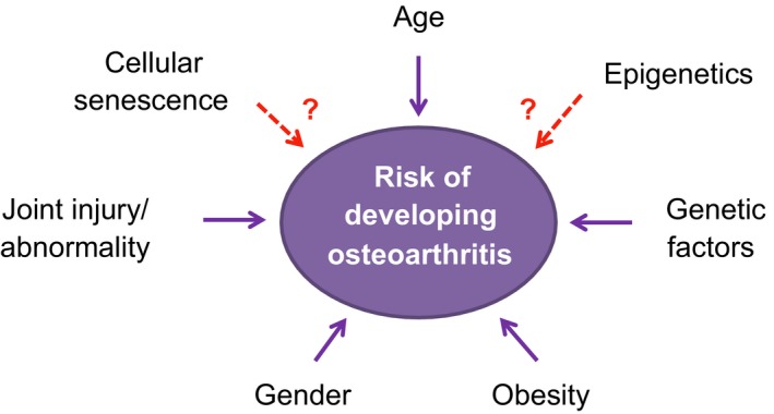
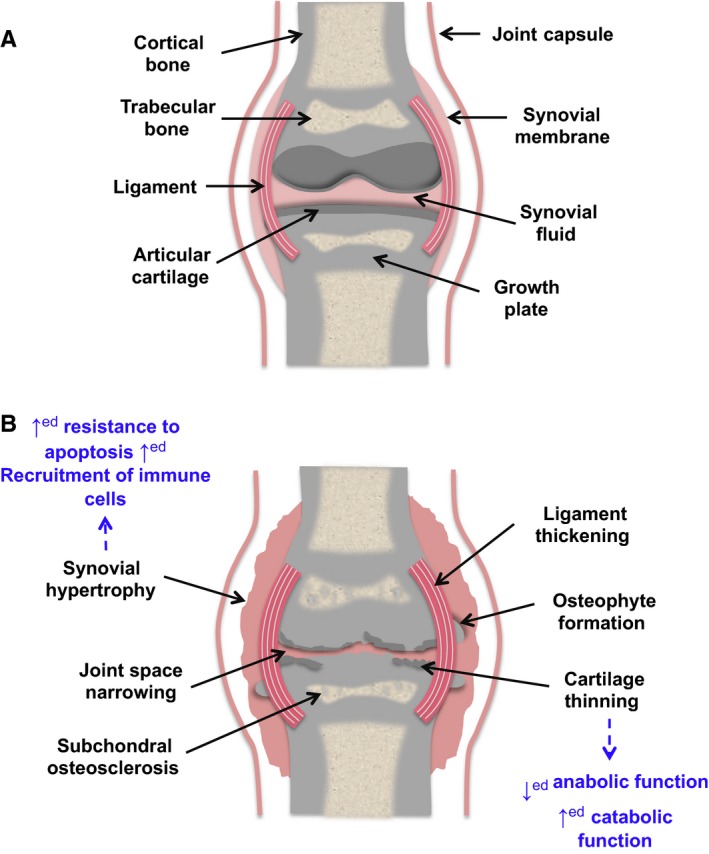
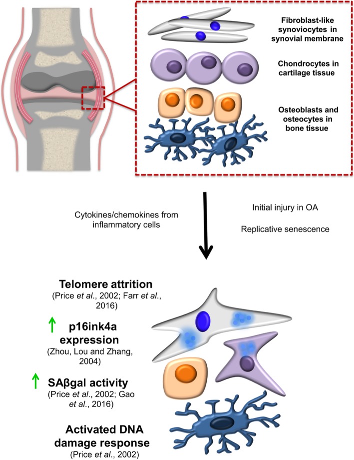
Cellular senescence is a state of stable proliferation arrest of cells. The senescence pathway has many beneficial effects and is seen to be activated in damaged/stressed cells, as well as during embryonic development and wound healing. However, the persistence and accumulation of senescent cells in various tissues can also impair function and have been implicated in the pathogenesis of many age-related diseases. Osteoarthritis (OA), a severely debilitating chronic condition characterized by progressive tissue remodeling and loss of joint function, is the most prevalent disease of the synovial joints, and increasing age is the primary OA risk factor. The profile of inflammatory and catabolic mediators present during the pathogenesis of OA is strikingly similar to the secretory profile observed in 'classical' senescent cells. During OA, chondrocytes (the sole cell type present within articular cartilage) exhibit increased levels of various senescence markers, such as senescence-associated beta-galactosidase (SAβGal) activity, telomere attrition, and accumulation of p16ink4a. This suggests the hypothesis that senescence of cells within joint tissues may play a pathological role in the causation of OA. In this review, we discuss the mechanisms by which senescent cells may predispose synovial joints to the development and/or progression of OA, as well as touching upon various epigenetic alterations associated with both OA and senescence.
Keywords: cellular senescence; epigenetics; osteoarthritis.
© 2017 The Authors. Aging Cell published by the Anatomical Society and John Wiley & Sons Ltd.
Figures
References
-
- Acosta JC, O'Loghlen A, Banito A, Guijarro MV, Augert A, Raguz S, Fumagalli M, Da Costa M, Brown C, Popov N, Takatsu Y, Melamed J, d'Adda di Fagagna F, Bernard D, Hernando E, Gil J (2008) Chemokine signaling via the CXCR2 receptor reinforces senescence. Cell 133, 1006–1018. - PubMed
-
- Acosta JC, Banito A, Wuestefeld T, Georgilis A, Janich P, Morton JP, Athineos D, Kang TW, Lasitschka F, Andrulis M, Pascual G, Morris KJ, Khan S, Jin H, Dharmalingam G, Snijders AP, Carroll T, Capper D, Pritchard C, Inman GJ, Longerich T, Sansom OJ, Benitah SA, Zender L, Gil J (2013) A complex secretory program orchestrated by the inflammasome controls paracrine senescence. Nat. Cell Biol. 15, 978–990. - PMC - PubMed
-
- d'Adda di Fagagna F (2008) Living on a break: cellular senescence as a DNA‐damage response. Nat. Rev. Cancer 8, 512–522. - PubMed
-
- d'Adda di Fagagna F, Reaper PM, Clay‐Farrace L, Fiegler H, Carr P, Von Zglinicki T, Saretzki G, Carter NP, Jackson SP (2003) A DNA damage checkpoint response in telomere‐initiated senescence. Nature 426, 194–198. - PubMed
-
- Aigner T, Hemmel M, Neureiter D, Gebhard PM, Zeiler G, Kirchner T, McKenna L (2001) Apoptotic cell death is not a widespread phenomenon in normal aging and osteoarthritis human articular knee cartilage: a study of proliferation, programmed cell death (apoptosis), and viability of chondrocytes in normal and osteoarthritic human knee cartilage. Arthritis Rheum. 44, 1304–1312. - PubMed
Publication types
MeSH terms
LinkOut - more resources
Full Text Sources
Other Literature Sources
Medical
