

Pannexin channel and connexin hemichannel expression in vascular function and inflammation
- PMID: 28124621
- PMCID: PMC5267334
- DOI: 10.1186/s12860-016-0119-3
Pannexin channel and connexin hemichannel expression in vascular function and inflammation
Abstract
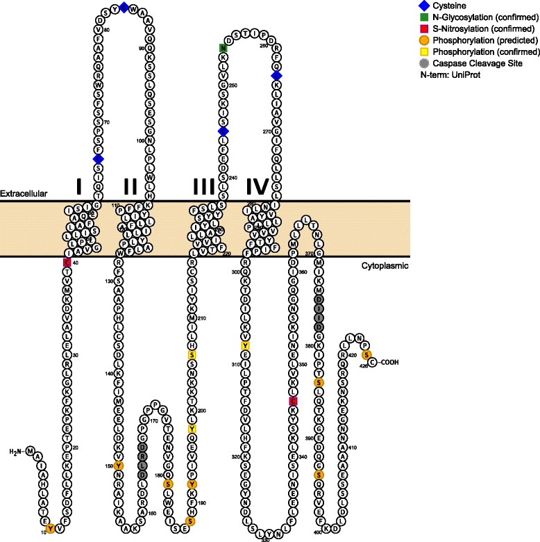
Control of blood flow distribution and tissue homeostasis depend on the tight regulation of and coordination between the microvascular network and circulating blood cells. Channels formed by connexins or pannexins that connect the intra- and extracellular compartments allow the release of paracrine signals, such as ATP and prostaglandins, and thus play a central role in achieving fine regulation and coordination of vascular function. This review focuses on vascular connexin hemichannels and pannexin channels. We review their expression pattern within the arterial and venous system with a special emphasis on how post-translational modifications by phosphorylation and S-nitrosylation of these channels modulate their function and contribute to vascular homeostasis. Furthermore, we highlight the contribution of these channels in smooth muscle cells and endothelial cells in the regulation of vasomotor tone as well as how these channels in endothelial cells regulate inflammatory responses such as during ischemic and hypoxic conditions. In addition, this review will touch on recent evidence implicating a role for these proteins in regulating red blood cell and platelet function.
Keywords: Connexins; Endothelium; Inflammation; Pannexins; Smooth muscle; Vasculature.
Figures
References
Publication types
MeSH terms
Substances
Grants and funding
LinkOut - more resources
Full Text Sources
Other Literature Sources
