Review
. 2017 Jan 17;18(Suppl 1):5.
doi: 10.1186/s12860-016-0121-9.

Accessing gap-junction channel structure-function relationships through molecular modeling and simulations

Affiliations
Review

Accessing gap-junction channel structure-function relationships through molecular modeling and simulations

F Villanelo et al. BMC Cell Biol. .

Abstract

Background: Gap junction channels (GJCs) are massive protein channels connecting the cytoplasm of adjacent cells. These channels allow intercellular transfer of molecules up to ~1 kDa, including water, ions and other metabolites. Unveiling structure-function relationships coded into the molecular architecture of these channels is necessary to gain insight on their vast biological function including electrical synapse, inflammation, development and tissular homeostasis. From early works, computational methods have been critical to analyze and interpret experimental observations. Upon the availability of crystallographic structures, molecular modeling and simulations have become a valuable tool to assess structure-function relationships in GJCs. Modeling different connexin isoforms, simulating the transport process, and exploring molecular variants, have provided new hypotheses and out-of-the-box approaches to the study of these important channels.

Methods: Here, we review foundational structural studies and recent developments on GJCs using molecular modeling and simulation techniques, highlighting the methods and the cross-talk with experimental evidence.

Results and discussion: By comparing results obtained by molecular modeling and simulations techniques with structural and functional information obtained from both recent literature and structural databases, we provide a critical assesment of structure-function relationships that can be obtained from the junction between theoretical and experimental evidence.

Keywords: Connexins; Gap-junction channels; Hemichannels; Homology modeling; Molecular simulation; Structure and function.


Figures
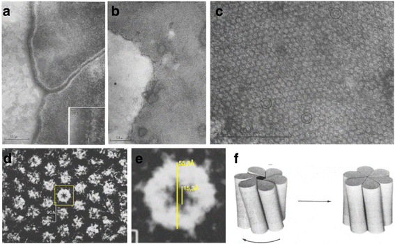
Fig. 1
Fig. 1
Early electron microscopy images of gap junction channels. a Cellular membrane between two rat liver cells. b Cellular membrane of rat liver cells immediately after isolation exhibiting the characteristic hexagonal pattern. c A highly magnified portion of the cellular membrane shown in Panel b. d Higher magnification and rotation of a portion of the cellular membrane shown in Panel c. e A digital zoom-in to the central yellow box denoting the extracellular portion of a hemichannel. Note the clear hexagonal symmetry. f The open/close model of a GJC proposed by Unwin and Zampighi [11]. Panels a to e, adapted from Benedetti and Emmelot [6]. Panel f, adapted from Unwin and Zampighi [11]
Fig. 2
Fig. 2
Early gap junction structures determined by electron crystallography and modelling. a A 3D EM-derived map of a Cx43 GJC. b The densities at different positions show clearly the 24 TMs, four for each monomer. c Model of Cα atoms derived by Fleishman et al., [21], showing in yellow the residues identified as pore lining. Panel a-b adapted from Unger et al., [16]; Panel c adapted from Fleishman et al., [21]
Fig. 3
Fig. 3
Schematic representation of the basic structural biology of connexins. a Secondary structure representation of a gap junction channel formed by end-to-end docking of two hemichannels of Cx26, colored in green and blue. Membrane planes depicted by red solid lines. b Representative hemichannel invoving hexameric arrangement of connexin protomers. Protomer domains appear denoted using color-coded names in one protomer as reference. c Intracellular view of a human Cx26 hemichannel in its open conformation with inner pore diameter of 14 Å (see text for explanation on channel openness). d 2D-plot denoting connexin secondary structure and the approximate position of every residue. The 3D coordinates of the human Cx26 gap junction and membrane planes were retrieved from Orientation of Proteins in Membrane Database [109] (using PDB id: 2ZW3). Panels a to c were rendered using Pymol. Panel d was modified from Nakagawa et al. [32]
Fig. 4
Fig. 4
Potential of Mean Force (PMF) as a function of pore length for permeating maltosaccharide solutes compared with pore radius. Taken from Luo et al., [90]
Fig. 5
Fig. 5
Schematic representation of a typical HC-GJC system simulated in MD. The HC is shown in cartoon representation in yellow. The dimensions are approximated, they could differ in different simulations. Simulation box is typically filled with solvent molecules, i.e. water and ions, not shown for simplicity. Figure prepared with software Pymol
Fig. 6
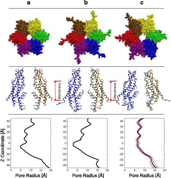
Fig. 6
Structure of hCx26. Column (a) crystallographic structure [19]; Column (b) modeled structure with completed CL and C-terminus plus missing residues and sidechains; Column (c), modeled structure after MD simulation. Upper row, structure of the channel viewed from extracellular side. Middle row,cartoon representation of two opposing monomers in a side view, with pore-lining residues in ball & stick. Lower row, pore radius as a function of pore length. Taken from Kwon et al., [22]
Fig. 7
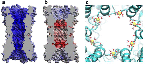
Fig. 7
Electrostatic potential surface on crystallographic structures of Cx26 (a) Ca2+ -bound and (b) Ca2+ -free. c Extracellular view of the interior of the Cx26 pore, highlighting the calcium binding site at the boundaries of the parahelix (PH). Taken from Bennett et al., [99]
Fig. 8
Fig. 8
The IC pocket of hCx26. a Schematics of IC pocket localization in one monomer, viewed from top. b Localization of the IC pocket between NTH, TM2, TM3, TM4 and TM1, denoting water inside the pocket using van der Waals representation. c Amino acid residues composing the IC pocket represented using sticks and colored by atom type. For clarity, the channel has been rotated 180° on the vertical axis with respect to (b) and the NTH removed. Taken from Araya-Secchi et al. [101]. Following the convention of Maeda et al. [19], the intracellular membrane face is located at top

References

    1. Goodenough DA. In vitro formation of gap junction vesicles. J Cell Biol. 1976;68:220–31. doi: 10.1083/jcb.68.2.220. - DOI - PMC - PubMed
    1. Harris AL. Emerging issues of connexin channels: biophysics fills the gap. Q Rev Biophys. 2001;34:325–472. doi: 10.1017/S0033583501003705. - DOI - PubMed
    1. Saez JC, Berthoud VM, Branes MC, Martinez AD, Beyer EC. Plasma membrane channels formed by connexins: their regulation and functions. Physiol Rev. 2003;83:1359–400. doi: 10.1152/physrev.00007.2003. - DOI - PubMed
    1. Robertson JD. The occurrence of a subunit pattern in the unit membranes of club endings in Mauthner cell synapses in goldfish brains. J Cell Biol. 1963;19:201–21. doi: 10.1083/jcb.19.1.201. - DOI - PMC - PubMed
    1. Robertson JD, Bodenheimer TS, Stage DE. The ultrastructure of Mauthner cell synapses nodes in goldfish. J Cell Biol. 1963;19:159–99. doi: 10.1083/jcb.19.1.159. - DOI - PMC - PubMed

Publication types

LinkOut - more resources