Review
. 2017 Jan 17;18(Suppl 1):6.
doi: 10.1186/s12860-016-0116-6.

Focus on lens connexins

Affiliations
Review

Focus on lens connexins

Viviana M Berthoud et al. BMC Cell Biol. .

Abstract

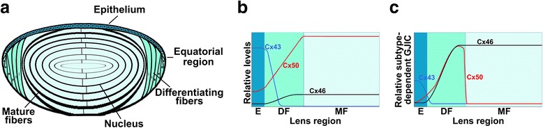
The lens is an avascular organ composed of an anterior epithelial cell layer and fiber cells that form the bulk of the organ. The lens expresses connexin43 (Cx43), connexin46 (Cx46) and connexin50 (Cx50). Epithelial Cx50 has critical roles in cell proliferation and differentiation, likely involving growth factor-dependent signaling pathways. Both Cx46 and Cx50 are crucial for lens transparency; mutations in their genes have been linked to congenital and age-related cataracts. Congenital cataract-associated connexin mutants can affect protein trafficking, stability and/or function, and the functional effects may differ between gap junction channels and hemichannels. Dominantly inherited cataracts may result from effects of the connexin mutant on its wild type isotype, the other co-expressed wild type connexin and/or its interaction with other cellular components.


Figures

Fig. 1
Fig. 1
Distribution, levels and contribution of lens connexins to gap junction coupling. a. Diagram showing the distribution of connexin subtypes in the different regions of the lens. b. Diagram showing the relative amounts of Cx43, Cx46 and Cx50 in the different lens regions of the adult mouse lens. The relative values of Cx43 and Cx50 in the epithelium are based on the quinine-sensitive component of intercellular communication between mouse lens epithelial cells [111]. The relative values of Cx46 and Cx50 in mature fibers are based on analysis of the membrane proteome of the mouse lens fiber cell [13]. The relative amounts on differentiating fibers cells have been drawn partially based on the up-regulation of Cx46 and downregulation of Cx43 expressions between epithelial and mature fiber cells, and the increase in Cx50 between epithelial and fiber cells as detected by immunofluorescence. c. Diagram showing the relative contributions of the different connexin subtypes to gap junction intercellular coupling in cells from the different regions of the adult mouse lens. The curves summarize the data from knockout and knock-in mouse experiments [–114]. The lens regions have been labeled E, epithelium; DF, differentiating fibers; MF, mature fibers. It is interesting to note that the contribution of Cx46 and Cx50 to gap junction coupling in fiber cells does not correlate with their protein levels
Fig. 2
Fig. 2
Docking of Cx46 connexons from adjacent cells. a. Homology model of the Cx46 gap junction channel based on the crystal structure of Cx26. The docking connexons are given in transparent surface representation. The interacting connexins of each connexon are shown as ribbons in gold and cyan, respectively. b. The network of hydrogen bonds between the amino acid residues of the interacting extracellular loops E1 and E2 are shown (dashed lines). (Reprinted with permission from [57])
Fig. 3
Fig. 3
Depiction of cataract mutants in human Cx46 and Cx50. Diagrams show the membrane topology of Cx46 and Cx50 and the localization of Cx46 and Cx50 mutants found in patients with non-syndromic and syndromic cataracts. The diagrams do not include mutations predisposing to age-related cataracts. Missense mutations are represented by a fusiform shape; frame-shift mutations (fs) are depicted by double rhombi; and early termination mutations by rectangles. Cx46 and Cx50 mutants with autosomal dominant inheritance are represented in light blue-purple. Mutants with recessive inheritance are shown in light golden yellow and mutants with an unassigned or sporadic inheritance pattern are shown in green. For different missense mutations at the same amino acid residue, the color coding depicts a known inheritance pattern (even if it is unassigned for the other missense mutations at that site). The Cx50I247M mutant has not been depicted, because it may be a polymorphism. These diagrams are based on the information available in Cat-Map ([2]; http://cat-map.wustl.edu/)
Fig. 4
Fig. 4
Interaction of fiber cell connexins with other lens components. The diagram depicts proteins which either interact with Cx46 and/or Cx50, or are affected by mutations in Cx46 or Cx50, or whose mutations affect Cx46 or Cx50 (as described in the text). The interaction of Cx50 with aquaporin0 is based on the report by Yu et al. [115]

References

    1. Goodenough DA. The crystalline lens. A system networked by gap junctional intercellular communication. Semin Cell Biol. 1992;3:49–58. doi: 10.1016/S1043-4682(10)80007-8. - DOI - PubMed
    1. Shiels A, Bennett TM, Hejtmancik JF. Cat-Map: putting cataract on the map. Mol Vis. 2010;16:2007–15. - PMC - PubMed
    1. Jiang JX. Gap junctions or hemichannel-dependent and independent roles of connexins in cataractogenesis and lens development. Curr Mol Med. 2010;10:851–63. doi: 10.2174/156652410793937750. - DOI - PMC - PubMed
    1. Rao PV. The pulling, pushing and fusing of lens fibers. Cell Adhes Migr. 2008;2:170–3. doi: 10.4161/cam.2.3.6495. - DOI - PMC - PubMed
    1. Beyer EC, Kistler J, Paul DL, Goodenough DA. Antisera directed against connexin43 peptides react with a 43-kD protein localized to gap junctions in myocardium and other tissues. J Cell Biol. 1989;108:595–605. doi: 10.1083/jcb.108.2.595. - DOI - PMC - PubMed

Publication types

Substances

LinkOut - more resources