. 2017 Jun 15;195(12):1576-1585.
doi: 10.1164/rccm.201608-1580OC.

Epstein-Barr Virus-induced Gene 2 Mediates Allergen-induced Leukocyte Migration into Airways

Affiliations

Epstein-Barr Virus-induced Gene 2 Mediates Allergen-induced Leukocyte Migration into Airways

Zhong-Jian Shen et al. Am J Respir Crit Care Med. .

Abstract

Rationale: Leukocyte recruitment to sites of allergic inflammation depends on the local production of priming cytokines, chemokines, and potentially other mediators. Previously, we showed that eosinophils (Eos) express numerous orphan G-protein-coupled receptors, including Epstein-Barr virus-induced gene 2 (EBI2). Despite its contribution to inflammatory diseases, the role of EBI2 in pulmonary eosinophilia is unknown.

Objectives: To determine whether oxysterol ligands for EBI2 are increased in asthma exacerbation, and if or how they promote Eos pulmonary migration.

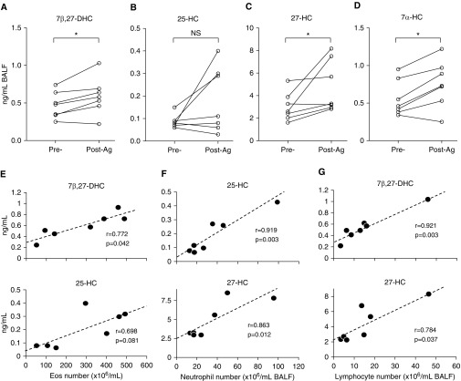
Methods: EBI2 ligands and pulmonary eosinophilia were measured in the bronchoalveolar lavage fluid from patients with mild asthma 48 hours after acute allergen challenge. In vitro, the ability of EBI2 ligands alone or in combination with IL-5 priming to induce the migration of human blood Eos was assessed.

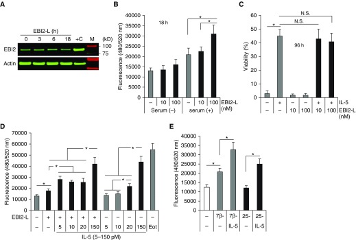
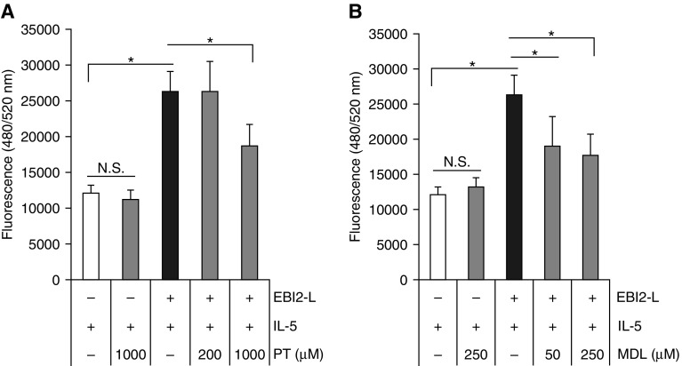
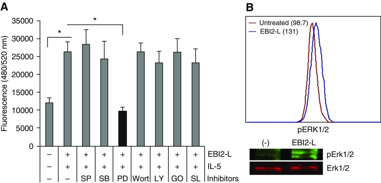
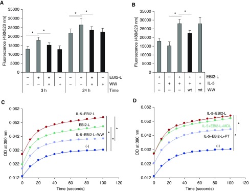
Measurements and main results: EBI2 was constitutively and stably expressed in peripheral blood Eos. Eos treated with the EBI2 ligands showed significantly increased transwell migration that was enhanced by priming with physiologic doses of IL-5. Migration was suppressed by inhibitors of the prolyl isomerase Pin1 or extracellular signal-regulated kinases (ERK) 1/2 or by pertussis toxin. EBI2 signaling activated Pin1 isomerase activity through a cascade that was sensitive to ERK inhibitors and pertussis toxin. The concentration of EBI2 ligands was significantly increased in the bronchoalveolar lavage fluid 48 hours after segmental allergen challenge and strongly correlated with the increased numbers of Eos, lymphocytes, and neutrophils in the airways.

Conclusions: Oxysterols are increased in inflamed airways after allergen challenge and, through G-protein subunit α, ERK, and Pin1 signaling, likely participate in the regulation of Eos migration into the lung in people with asthma.

Keywords: EBI2; Pin1; asthma; eosinophils; migration.


Figures

Figure 1.
Figure 1.
Epstein-Barr virus–induced gene 2 ligands are increased in bronchoalveolar lavage fluid (BALF) after segmental allergen challenge. (A–D) BALF was collected before and 48 hours after segmental allergen challenge of patients (n = 7) with mild asthma. After removal of cells, the supernatants were subjected to mass spectrometry. (E–G) Pearson correlation coefficients (r) were measured to evaluate the linear relationship between ligand concentration and cell numbers in BAL from after segmental allergen challenge. *P < 0.05 by paired Student's t test. Ag = allergen challenge; DHC = dihydroxycholesterol; Eos = eosinophil; HC = hydroxycholesterol; NS = nonsignificant.
Figure 2.
Figure 2.
Epstein-Barr virus–induced gene 2 (EBI2) is expressed by eosinophil (Eos) and directs migration. (A) Freshly purified blood Eos were treated with EBI2 ligand (EBI2-L) (7α, 25-dihydroxycholesterol [DHC]) (100 nM = 40.3 ng/ml) for the indicated times. Whole-cell lysates were subjected to immunoblots with anti-EBI2 and β-actin as shown. +C: Ramos cell lysate served as positive control. (B) Cell migration assay was performed (see Methods) in the presence (+) or absence (−) of 10% fetal bovine serum and EBI2-L. Migrated cells were lysed and stained with DNA dye. (C) Cells were cultured for 96 hours in the presence or absence of EBI2-L (10–100 nM) and IL-5 (150 pM) in the medium containing 10% fetal bovine serum before viability assay. (D) Cells were primed in the presence of different doses of IL-5 (5–150 pM) for 10 minutes before induction of migration with EBI2-L (100 nM) for 3 hours. Eotaxin (50 ng/ml) used as a positive control. (E) Cells were primed with IL-5 (10 pM) before induction of migration with other EBI2-L (6 μM of 7β,27-DHC, or 152 μM of 25-DHC) for 3 hours. Data are expressed as mean ± SD. *P < 0.05. The data are from at least four independent experiments. Eot = eotaxin; N.S. = nonsignificant.
Figure 3.
Figure 3.
G-protein α and cAMP mediate Epstein-Barr virus–induced gene 2 (EBI2) signaling. (A and B) Eosinophils were pretreated for 10 minutes with pertussis toxin or adenylyl cyclase inhibitor (MDL-12330A) before priming with IL-5 (10 pM, 10 min) and subsequent induction of migration with EBI2 ligand (100 nM) for 3 hours. *P < 0.05. The data are from at least four independent experiments. EBI2-L = Epstein-Barr-virus-induced gene 2 ligand; MDL = MDL-12330A; N.S. = nonsignificant; PT = pertussis toxin.
Figure 4.
Figure 4.
Extracellular signal–regulated kinase (ERK) mitogen-activated protein kinase (MAPK) mediates Epstein-Barr virus–induced gene 2 (EBI2) signaling. (A) Eosinophils were pretreated with kinase inhibitors (5 μM SP600125 [SP] for c-Jun N-terminal kinases, 5 μM SB203580 [SB] for p38 MAPK, 50 μM PD98059 [PD] for ERK MAPK, 200 nM wortmannin [Wort] for phosphatidylinositol-3 kinases, 10 μM LY294002 [LY] for phosphatidylinositol-3 kinases, 10 nM Gö6976 [GO] for protein kinase C-α/β, 100 nM SL0101-1 [SL] for p90 ribosomal S6 kinase) before priming with IL-5 (10 pM) and subsequent induction of migration with EBI2 ligand (EBI2-L) (100 nM) for 3 hours. (B) Cells were treated with EBI2-L alone for 10 minutes and subjected to flow cytometry and immunoblots for the activation of ERK1/2. *P < 0.05. The data are from at least four independent experiments.
Figure 5.
Figure 5.
Pin1 is required for Epstein-Barr virus–induced gene 2 (EBI2)-directed eosinophil migration. (A) Eosinophils were pretreated with dominant negative TAT-WW of Pin1 peptide (WW, 10 nM) for 10 minutes before induction of migration with EBI2 ligand (EBI2-L) (100 nM) alone for the indicated times. (B) Cells were pretreated with TAT-WW (wt) or with its nonfunctional mutant (mt) before priming with IL-5 (10 pM) and subsequent induction of migration with EBI2 ligand for 3 hours. (C and D) Cells were treated as in B for 10 minutes. Cell lysates were subjected to Pin1 isomerase activity assay as described in Methods. In D, 1,000 μM of pertussis toxin and 250 μM of MDL were used. *P < 0.05. The data are from at least four independent experiments. MDL = MDL-12330A; OD = optical density; PT = pertussis toxin.

Comment in

References

    1. Rothenberg ME, Hogan SP. The eosinophil. Annu Rev Immunol. 2006;24:147–174. - PubMed
    1. Humbles AA, Lu B, Friend DS, Okinaga S, Lora J, Al-Garawi A, Martin TR, Gerard NP, Gerard C. The murine CCR3 receptor regulates both the role of eosinophils and mast cells in allergen-induced airway inflammation and hyperresponsiveness. Proc Natl Acad Sci USA. 2002;99:1479–1484. - PMC - PubMed
    1. Gauvreau GM, Boulet LP, Cockcroft DW, Baatjes A, Cote J, Deschesnes F, Davis B, Strinich T, Howie K, Duong M, et al. Antisense therapy against CCR3 and the common beta chain attenuates allergen-induced eosinophilic responses. Am J Respir Crit Care Med. 2008;177:952–958. - PubMed
    1. Neighbour H, Boulet LP, Lemiere C, Sehmi R, Leigh R, Sousa AR, Martin J, Dallow N, Gilbert J, Allen A, et al. Safety and efficacy of an oral CCR3 antagonist in patients with asthma and eosinophilic bronchitis: a randomized, placebo-controlled clinical trial. Clin Exp Allergy. 2014;44:508–516. - PubMed
    1. Nagase H, Kudo K, Izumi S, Ohta K, Kobayashi N, Yamaguchi M, Matsushima K, Morita Y, Yamamoto K, Hirai K. Chemokine receptor expression profile of eosinophils at inflamed tissue sites: decreased CCR3 and increased CXCR4 expression by lung eosinophils. J Allergy Clin Immunol. 2001;108:563–569. - PubMed

Publication types