

A Feasibility Study of Quantifying Longitudinal Brain Changes in Herpes Simplex Virus (HSV) Encephalitis Using Magnetic Resonance Imaging (MRI) and Stereology
- PMID: 28125598
- PMCID: PMC5268482
- DOI: 10.1371/journal.pone.0170215
A Feasibility Study of Quantifying Longitudinal Brain Changes in Herpes Simplex Virus (HSV) Encephalitis Using Magnetic Resonance Imaging (MRI) and Stereology
Abstract
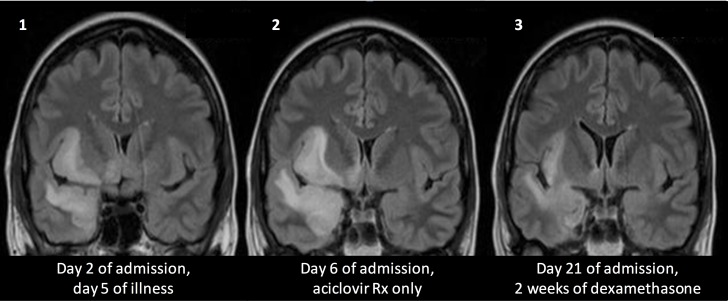
Objectives: To assess whether it is feasible to quantify acute change in temporal lobe volume and total oedema volumes in herpes simplex virus (HSV) encephalitis as a preliminary to a trial of corticosteroid therapy.
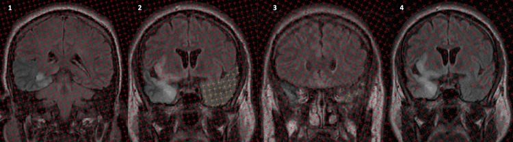
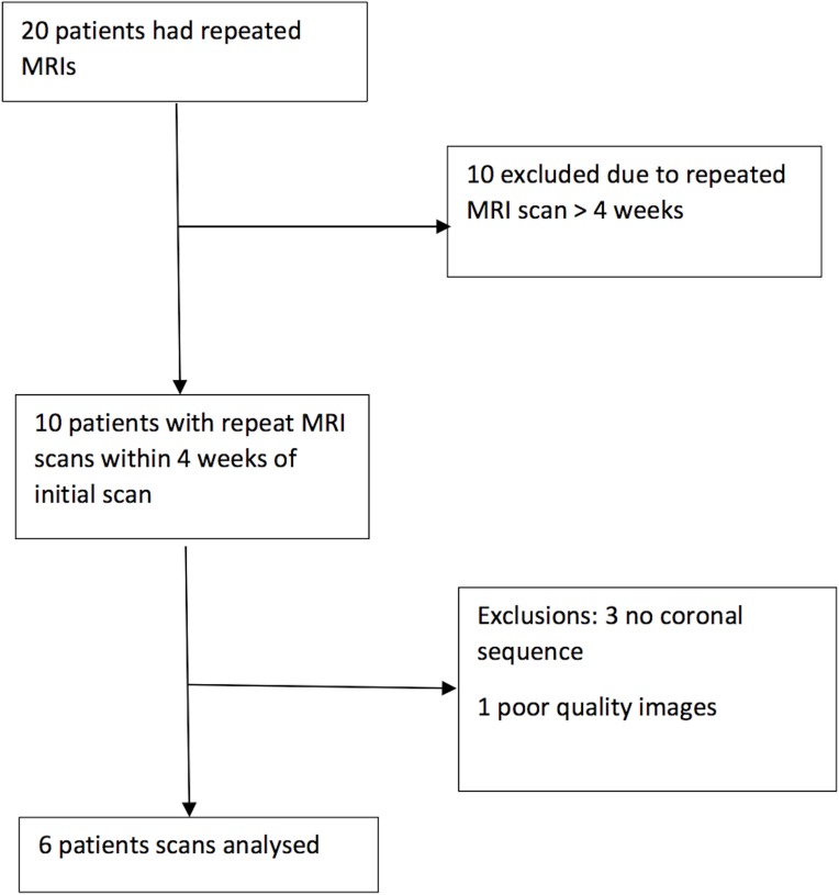
Methods: The study analysed serially acquired magnetic resonance images (MRI), of patients with acute HSV encephalitis who had neuroimaging repeated within four weeks of the first scan. We performed volumetric measurements of the left and right temporal lobes and of cerebral oedema visible on T2 weighted Fluid Attenuated Inversion Recovery (FLAIR) images using stereology in conjunction with point counting.
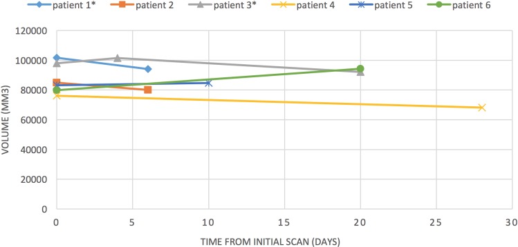
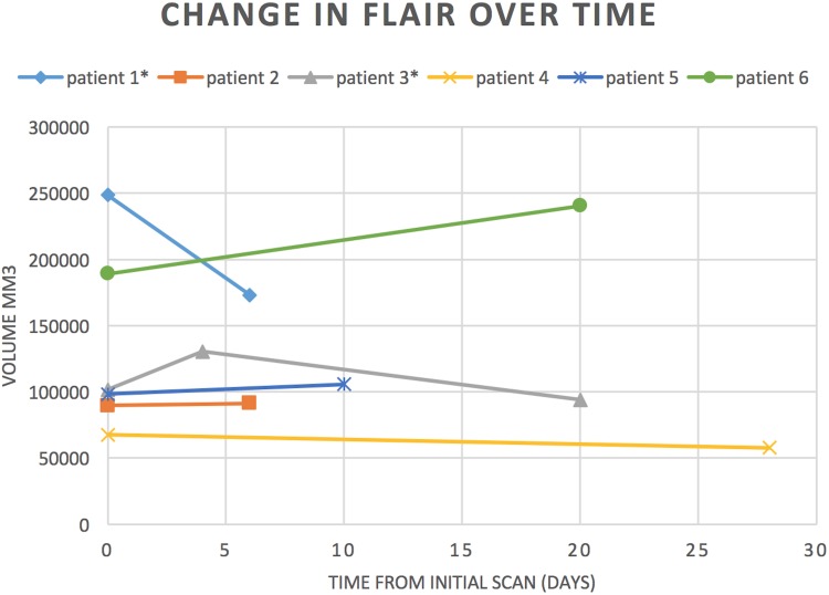
Results: Temporal lobe volumes increased on average by 1.6% (standard deviation (SD 11%) in five patients who had not received corticosteroid therapy and decreased in two patients who had received corticosteroids by 8.5%. FLAIR hyperintensity volumes increased by 9% in patients not receiving treatment with corticosteroids and decreased by 29% in the two patients that had received corticosteroids.
Conclusions: This study has shown it is feasible to quantify acute change in temporal lobe and total oedema volumes in HSV encephalitis and suggests a potential resolution of swelling in response to corticosteroid therapy. These techniques could be used as part of a randomized control trial to investigate the efficacy of corticosteroids for treating HSV encephalitis in conjunction with assessing clinical outcomes and could be of potential value in helping to predict the clinical outcomes of patients with HSV encephalitis.
Conflict of interest statement
Dr Simon S Keller is funded by a UK Medical Research Council grant, grant number MR/K023152/1 and Prof Tom Solomon is funded by the National Institute for Health Research by a Programme Grant for Applied Research with grant number RP-PG-108-10048. All other authors have no competing interests.
Figures
References
Publication types
MeSH terms
Substances
Grants and funding
LinkOut - more resources
Full Text Sources
Other Literature Sources
Medical
