. 2017 Jan 26;12(1):e0170305.
doi: 10.1371/journal.pone.0170305. eCollection 2017.

A Recombinant Human Anti-Platelet scFv Antibody Produced in Pichia pastoris for Atheroma Targeting

Affiliations

A Recombinant Human Anti-Platelet scFv Antibody Produced in Pichia pastoris for Atheroma Targeting

Amelie Vallet-Courbin et al. PLoS One. .

Abstract

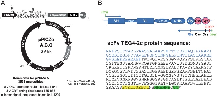
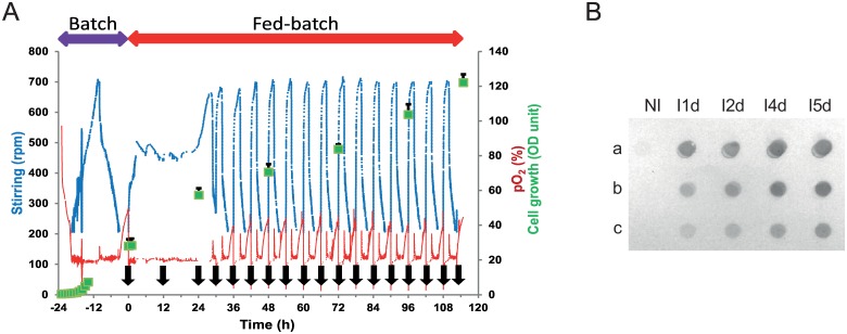
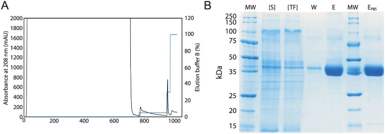
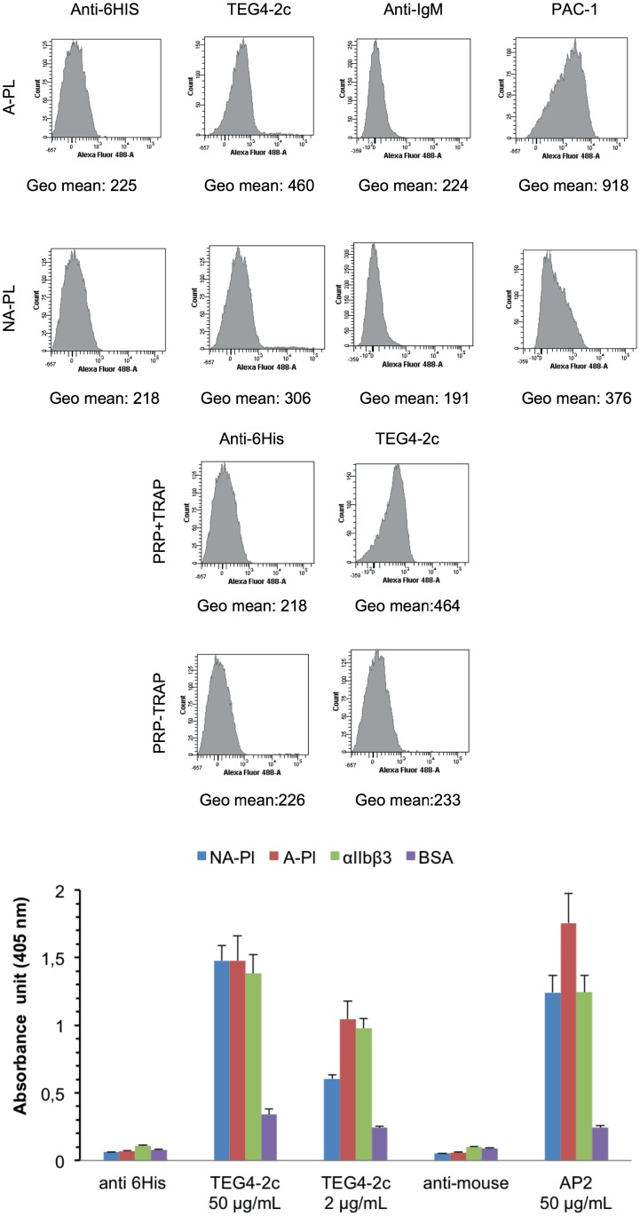
Cells of the innate and adaptive immune system are key factors in the progression of atherosclerotic plaque, leading to plaque instability and rupture, potentially resulting in acute atherothrombotic events such as coronary artery disease, cerebrovascular disease and peripheral arterial disease. Here, we describe the cloning, expression, purification, and immunoreactivity assessment of a recombinant single-chain variable fragment (scFv) derived from a human anti-αIIbβ3 antibody (HuAb) selected to target atheromatous lesions for the presence of platelets. Indeed, platelets within atheroma plaques have been shown to play a role in inflammation, in platelet-leucocyte aggregates and in thrombi formation and might thus be considered relevant biomarkers of atherosclerotic progression. The DNA sequence that encodes the anti-αIIbβ3 TEG4 scFv previously obtained from a phage-display selection on activated platelets, was inserted into the eukaryote vector (pPICZαA) in fusion with a tag sequence encoding 2 cysteines useable for specific probes grafting experiments. The recombinant protein was expressed at high yields in Pichia pastoris (30 mg/L culture). The advantage of P. pastoris as an expression system is the production and secretion of recombinant proteins in the supernatant, ruling out the difficulties encountered when scFv are produced in the cytoplasm of bacteria (low yield, low solubility and reduced affinity). The improved conditions allowed for the recovery of highly purified and biologically active scFv fragments ready to be grafted in a site-directed way to nanoparticles for the imaging of atherosclerotic plaques involving inflammatory processes and thus at high risk of instability.


Conflict of interest statement

Pall Life Sciences provided support on the Octet Red experiments. There are no patents, products in development or marketed products to declare. This does not alter the authors' adherence to all the PLOS ONE policies on sharing data and materials.

Figures

Fig 1
Fig 1. pPICZαA expression vector according to the EasySelect Pichia Expression Kit Manual (Invitrogen) and a schematic representation of TEG4-2c scFv with the protein sequence.
(A): All the featured restriction sites are unique. 5´ AOX1: promoter region of AOX1; TT AOX1: transcription termination of AOX1; PTEF1: promoter of TEF1; PEM7: promoter of EM7; Zeocin resistance: Sh ble ORF; CYC1 TT: transcription termination of CYC1. (B): The TEG4-2c scFv coding sequence was cloned between PmlI and XbaI sites and the protein sequence of recombinant tag-scFv including the 6His-tag and the 2 cysteine are highlighted in green. The α-factor signal sequence is represented in blue and the C-myc tag is highlighted in yellow.
Fig 2
Fig 2. TEG4-2c scFv production process. A: Fed batch fermentation history plot.
Stirring, pO2 and OD600 values are plotted versus time during the cultivation of P. pastoris in BMGY medium. Cultures were induced with methanol at t = 0 (24 h after starting the batch phase) during the fed batch phase the methanol was added every 12 h or 6h (black arrows) to a final concentration of 0.6%. The average values are shown with error bars representing the standard deviation (n = 5). 1 OD600 unit was equivalent to 0.29 mg/mL dry weight. B: Dot-blot analysis of supernatants from recombinant P. pastoris culture. Fifty microliters samples from non-induced culture (NI) and from day 1 to day 5 induced cultures (I1d to I5d) were undiluted (a) or diluted (b = 1:10; c = 1:50) and blotted on a nitrocellulose membrane. The recombinant TEG4-2c scFv were detected with the Anti-6His antibody and revealed by colorimetric analysis.
Fig 3
Fig 3
Purification of recombinant TEG4-2c scFv A: IMAC chromatogram. The HisTrap Excel resin (5 mL) was equilibrated with 50 mM Tris-HCl pH 7.5, 500 mM NaCl (buffer A at a flow rate of 3 mL/min). Pichia pastoris expression broth supernatant containing the TEG4-2c scFv was injected into the column. The column was then washed with buffer A until absorbance at 280nm reached the baseline. (Dot Line): The elution was carried out in two steps using 5% and 30% buffer B (50 mM Tris-HCl pH 7.5, 500 mM NaCl, 500 mM imidazole) corresponding respectively to 25 mM and 150 mM imidazole. B: Electrophoretic analysis of one step IMAC purification of recombinant TEG4-2c scFv. 12% SDS-PAGE stained with colloidal blue, MW: molecular weight ladder (KDa). [S]: 5x concentrated culture supernatant. [TF]: 5x concentrated flow-through. W: 25 mM imidazole washing fraction. E: 150 mM elution fraction. EPBS: Elution fraction dialyzed against PBS.
Fig 4
Fig 4. Binding assessment of TEG4-2c scFv to human platelets by flow cytometry and ELISA tests.
A: Binding of TEG4-2c scFv on thrombin-activated human (A-PL) or non-activated—platelets (NA-PL) analysed by flow cytometry. PAC-1 IgM murine antibody serves as a positive control. Binding of antibody to the platelets was further detected by incubation with Alexa Fluor 488 anti-6His or anti-mouse IgM antibodies. Negative controls were secondary antibody only. Histograms depict representative data ± SD of three independent experiments. Quantitative fluorescence intensities (in Geo mean) are stated under each respective histogram. B: Binding of TEG4-2c scFv on TRAP-activated-human (+ TRAP) or non-activated platelets (-TRAP) analysed by flow cytometry. Quantitative fluorescence intensities (in Geo mean) are stated under each respective histogram. C: Representative whole cell (A-PL, NA-PL) and purified proteins (αIIbβ3, BSA) ELISA with TEG4-2c scFv. A murine anti-αIIbβ3 antibody AP2 was used as positive control. Negative controls were secondary antibody only. Binding of antibodies was visualized via HRP-6His or HRP-anti-mouse IgG. OD value represents absorbance at 450 nm. Plots represent the mean values ± SD (n = 3)
Fig 5
Fig 5. Binding of scFv TEG4-2c to αIIbβ3 by SPR and to whole platelets by BLI.
A: SPR sensorgrams. The ligand αIIbβ3 was immobilized on CM5 chip by amine coupling with a density of 8000 RU. Serial dilutions of TEG4-2c in HBS-EP running buffer were injected over the ligand corresponding to 94, 188 and 390 nM. Sensorgrams show a binding concentration-dependent of TEG4-2c scFv. B: BLI analysis. TEG4-2c scFv (ligand) was loaded on HIS2 biosensor (anti-penta Histidine Ab) at 21 μg/mL. Whole platelets (analyte) concentrations converted into αIIbβ3 molarities were: 41.5, 8.3, 4.15, 0.83, 0.415, 0.083 and 0.0415 nM. Additionally one sensor pair was used to record the buffer reference signals. TEG4-2c scFv reacts with αIIbβ3 in its natural conformation in a concentration dependent manner.
Fig 6
Fig 6. Comparison of the immunoreactivity of TEG4-2c scFv to atherosclerotic tissues of different species by IHC analysis and ELISA assays.
A (a-r): IHC assays on atherosclerotic tissues: similarly to positive controls e.g; anti-CD41 (anti-αIIb) (e), RAM11 and PGM1 (anti-CD68 antibodies targeting rabbit and human macrophages respectively) (k, q) and AP2 (anti-αIIbβ3 antibody) (a; g; m), TEG4-2c scFv specifically recognizes the injured areas of the aorta sections from different species (c; i; o). Binding of antibodies was visualized via HRP-anti-6His (scFv); HRP anti-rabbit IgG (CD41) and HRP anti-mouse IgG (RAM11, AP2). Negative controls were secondary antibody only (b; d; f; h; j; l; n; p; r). Nuclei were counterstained with hematoxylin B: ELISA tests on atheromatous and healthy aorta proteins: TEG4-2c shows a better recognition of atheromatous proteins. RAM11 and AP2 were used as positive controls. Negative controls were secondary antibody only. Binding of antibodies was visualized via HRP-6His or HRP-anti-mouse IgG. OD value represents absorbance at 450 nm. Values represent mean (n = 3) ± SD (error bars materialized the SD)

Similar articles

Cited by

References

    1. Libby P, Ridker PM, Hansson GK (2011) Progress and challenges in translating the biology of atherosclerosis. Nature 473: 317–325. 10.1038/nature10146 - DOI - PubMed
    1. Falk E (2006) Pathogenesis of atherosclerosis. J Am Coll Cardiol 47: C7–12. 10.1016/j.jacc.2005.09.068 - DOI - PubMed
    1. Joshi FR, Lindsay AC, Obaid DR, Falk E, Rudd JH (2012) Non-invasive imaging of atherosclerosis. Eur Heart J Cardiovasc Imaging 13: 205–218. 10.1093/ehjci/jer319 - DOI - PubMed
    1. Wildgruber M, Swirski FK, Zernecke A (2013) Molecular imaging of inflammation in atherosclerosis. Theranostics 3: 865–884. 10.7150/thno.5771 - DOI - PMC - PubMed
    1. Weber C, Zernecke A, Libby P (2008) The multifaceted contributions of leukocyte subsets to atherosclerosis: lessons from mouse models. Nat Rev Immunol 8: 802–815. 10.1038/nri2415 - DOI - PubMed

MeSH terms

Substances

LinkOut - more resources