. 2017 Jan 26;13(1):e1006126.
doi: 10.1371/journal.ppat.1006126. eCollection 2017 Jan.

Toxoplasma gondii GRA7-Targeted ASC and PLD1 Promote Antibacterial Host Defense via PKCα

Affiliations

Toxoplasma gondii GRA7-Targeted ASC and PLD1 Promote Antibacterial Host Defense via PKCα

Hyun-Jung Koh et al. PLoS Pathog. .

Abstract

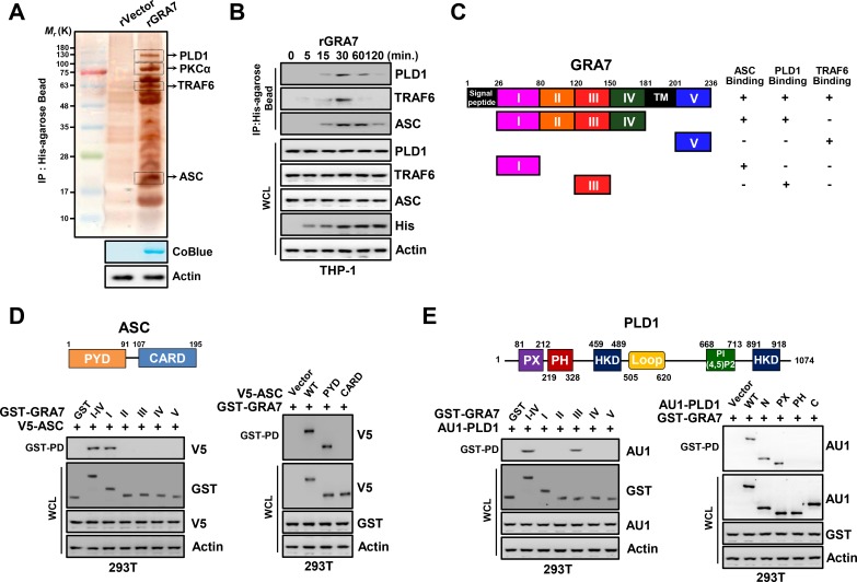
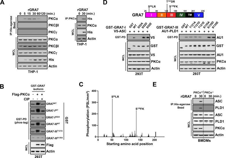
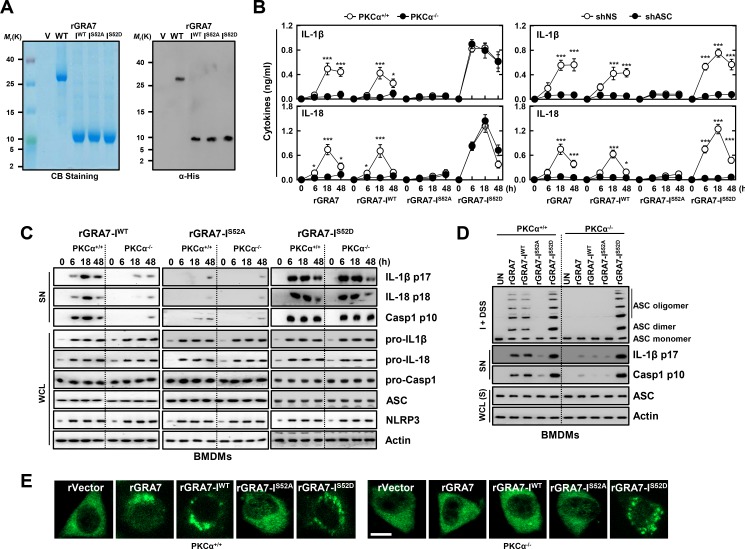
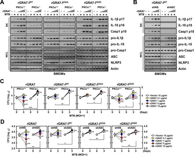
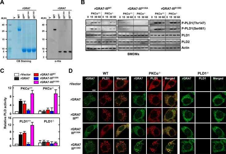
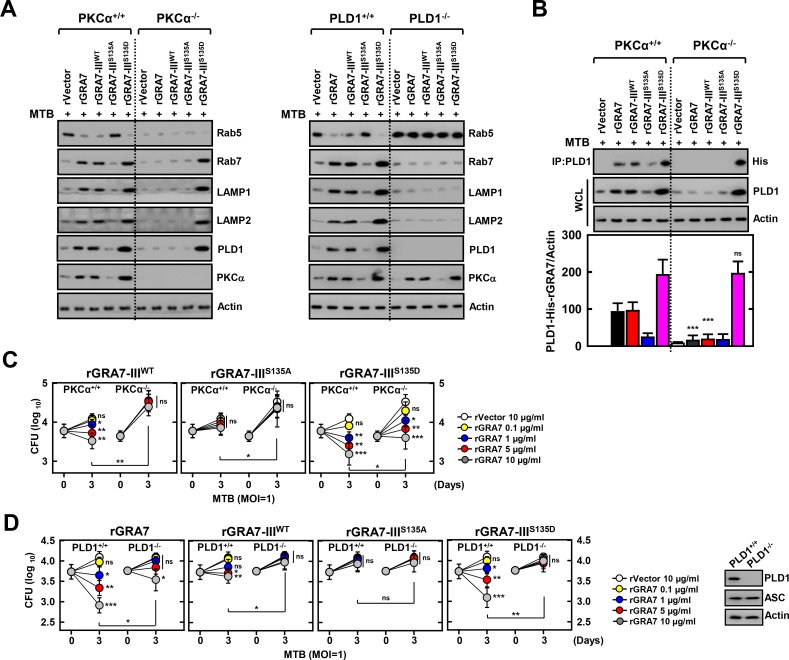
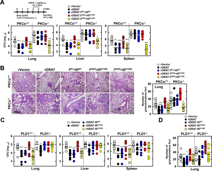
Tuberculosis is a global health problem and at least one-third of the world's population is infected with Mycobacterium tuberculosis (MTB). MTB is a successful pathogen that enhances its own intracellular survival by inhibiting inflammation and arresting phago-lysosomal fusion. We previously demonstrated that Toxoplasma gondii (T. gondii) dense granule antigen (GRA) 7 interacts with TNF receptor-associated factor 6 via Myeloid differentiation primary response gene 88, enabling innate immune responses in macrophages. To extend these studies, we found that GRA7 interacts with host proteins involved in antimicrobial host defense mechanisms as a therapeutic strategy for tuberculosis. Here, we show that protein kinase C (PKC)α-mediated phosphorylation of T. gondii GRA7-I (Ser52) regulates the interaction of GRA7 with PYD domain of apoptosis-associated speck-like protein containing a carboxy-terminal CARD, which is capable of oligomerization and inflammasome activation can lead to antimicrobial defense against MTB. Furthermore, GRA7-III interacted with the PX domain of phospholipase D1, facilitating its enzyme activity, phago-lysosomal maturation, and subsequent antimicrobial activity in a GRA7-III (Ser135) phosphorylation-dependent manner via PKCα. Taken together, these results underscore a previously unrecognized role of GRA7 in modulating antimicrobial host defense mechanism during mycobacterial infection.


Conflict of interest statement

The authors have declared that no competing interests exist.

Figures

Fig 1
Fig 1. GRA7 interactions with ASC and PLD1.
(A) Identification of PLD1, PKCα, TRAF6, and ASC by mass spectrometry analysis in THP-1 cell lysates incubated with a His-tagged rGRA7 (2 μg). CoBlue, staining of His-rGRA7 with Coomassie blue. Whole cell lysates (WCLs) were used for immuno blotting (IB) with αActin. (B) THP-1 cells were stimulated with rGRA7 (5 μg/ml) for the indicated times, followed by immunoprecipitation (IP) with αHis-agarose bead and IB with αPLD1, αTRAF6, αASC, αHis, and αActin. (C) Summary of the interactions of GRA7 WT and its mutants with ASC, PLD1, and TRAF6. The binding activities of GRA7 WT and mutants are summarized based on the results of Fig 1D and 1E, S2E Fig, and [17]. (D and E) Binding mapping. Schematic diagram of the structures of ASC and PLD1 (upper). (D) At 48 hr post-transfection with mammalian GST or GST-GRA7 and truncated mutant constructs together with V5-ASC (left), V5, or V5-ASC constructs together with GST-GRA7 (right), 293T cells were used for GST pulldown, followed by IB with αV5. WCLs were used for IB with αGST, αV5 or αActin. (E) 293T cells were co-transfected with GST or GST-GRA7 and truncated mutant constructs together with AU1-PLD1 (left), or AU1 or AU1-PLD1 constructs together with GST-GRA7 (right), and subjected to GST pulldown, followed by IB with αAU1. WCLs were used for IB with αGST, αAU1 or αActin. The data are representative of four independent experiments with similar results (A, B, D, E).
Fig 2
Fig 2. PKCα-dependent phosphorylation of GRA7 was essential for interaction with ASC and PLD1.
(A) THP-1 cells were stimulated with rGRA7 (5 μg/ml) for the indicated times (left) or 30 min. (right), followed by IP with αHis-agarose bead (left) or αPKCα (right) and IB with αPKCα, αPKCβI, αPKCγ, αHis, and αActin. (B) Phos-tag and SDS-PAGE analyses of GST-GRA7, GRA7-IWT, GRA7-IS52A, GRA7-IIIWT, GRA7-IIIT121A, or GRA7-IIIS135A expressed together with Flag-tagged PKCα in 293T cells left untreated (CIP-) or treated with calf intestinal alkaline phosphatase (CIP+), and subjected to GST pulldown, followed by IB with αGST. WCLs were used for IB with αFlag or αActin. (C) Mapping of PKCα phosphorylation sites on GRA7 by tiled peptide array analysis using purified recombinant PKCα. Phosphorylation intensity of 15-amino acid peptides that span full-length GRA7 and are each shifted by 3 amino acids was detected using MultiGauge version 3.0. The serines in the two peptides that showed a phosphorylation signal stronger than 100 PSL/mm2 are indicated above the corresponding peaks. (D) 293T cells were co-transfected with GST or GST-GRA7-I and truncated mutant constructs together with V5-ASC (left), or GST or GST-GRA7-III and truncated mutant constructs together with AU1-PLD1 (right), and subjected to GST pulldown, followed by IB with αV5 or αAU1. WCLs were used for IB with αGST, αV5, αAU1, αPKCα, or αActin. (E) BMDMs from PKCα+/+ and PKCα-/- were stimulated with rGRA7 for 30 min., followed by IP with αHis-agarose bead and IB with αASC, αPLD1, αPKCα, and αActin. The data are representative of four independent experiments with similar results (A to E).
Fig 3
Fig 3. GRA7-I activated inflammasomes in an ASC-binding dependent manner.
(A) Bacterially purified 6xHis-GRA7-I and its mutants were analyzed by Coomassie blue staining (left) or IB with αHis (right). (B) BMDMs from PKCα+/+ and PKCα-/- (left) or BMDMs were transduced with lentivirus-shRNA-NS or lentivirus-shRNA-ASC (MOI = 100) with polybrene (8 μg/mL) (right) for 2 days, the cells were stimulated with rGRA7 (5 μg/ml) and its mutants for the indicated times and culture supernatants were harvested and analyzed for cytokine ELISA for IL-1β and IL-18. Data shown are the means ± SD of five experiments. Significant differences (*P < 0.05; ***P < 0.001) compared with PKCα+/+ or shRNA-NS. (C) IB analysis for IL-1β p17, IL-18 p18, or caspase-1 p10 in supernatants (SN), ASC, NLRP3, pro-IL-1β, pro-IL-18, or pro-caspase-1 in whole-cell lysates (WCL). Actin was used as a loading control. (D) IB analysis of lysates of BMDMs as in B and C solubilized with Triton X-100–containing buffer, followed by cross-linkage of insoluble fractions with disuccinimidyl suberate to capture ASC oligomers and analysis of those fractions (I + DSS) and soluble fractions (S) with antibody to ASC. Actin was used as a loading control. (E) Fluorescence confocal images showing formation of speck-like ASC pyroptosomes in BMDMs from PKCα+/+ and PKCα-/- mice were stimulated with rGRA7 and its mutants for 18 h, fixed, immunostained with antibodies for ASC (Alexa 488). Scale bar, 10 μm. The data are representative of five independent experiments with similar results (A and C-E).
Fig 4
Fig 4. GRA7-I-induced inflammasome activation was required for antimicrobial activity in MTB-infected macrophages.
(A and B) BMDMs from PKCα+/+ and PKCα-/- mice (A) and BMDMs were transduced with lentivirus-shRNA-NS or lentivirus-shRNA-ASC for 2 days (B) infected with MTB (MOI = 1) for 4 h and then stimulated with rGRA7 (1, 5, 10 μg/ml) and its mutants for 18 h. IB analysis for IL-1β p17, IL-18 p18, or caspase-1 p10 in supernatants (SN), ASC, NLRP3, pro-IL-1β, pro-IL-18, or pro-caspase-1 in whole-cell lysates (WCL). Actin was used as a loading control. (C and D) Intracellular survival of MTB was assessed by CFU assay. BMDMs were infected with MTB for 4 h, followed by treatment with rGRA7, and then lysed to determine intracellular bacterial loads. The data are representative of five independent experiments with similar results (A and B). Data shown are the mean ± SD of five experiments (C and D). Significant differences (*P < 0.05; **P < 0.01; ***P < 0.001) compared with rVector. CFU, colony-forming units. ns, not significant.
Fig 5
Fig 5. GRA7-III-induced activation of PLD1 was dependent on interaction with PLD1.
(A) Bacterially purified 6xHis-GRA7-III and its mutants were analyzed by Coomassie blue staining (left) or IB with αHis (right). (B) BMDMs from PKCα+/+ and PKCα-/- mice were stimulated with rGRA7-III (5 μg/ml) and its mutants for the indicated times, followed by IB to detect phosphorylated and total forms of PLD1 and PLD2. Actin was used as a loading control. (C) BMDMs were stimulated with rGRA7-III and its mutants for 30 min. and analyzed for PLD activity assay in vitro. Data shown are the means ± SD of five experiments. (D) Fluorescence confocal images in BMDMs were stimulated with rGRA7 and its mutants for 30 min., fixed, immunostained with antibodies for His (Alexa 488) and PLD1 (Alexa 568). Scale bar, 20 μm. The data are representative of three independent experiments with similar results (A, B, and D).
Fig 6
Fig 6. GRA7-III-induced activation of PLD1 and phagosomal maturation were required for antimicrobial activity in MTB-infected macrophages.
(A and B) BMDMs were infected with MTB (MOI = 1) for 4 h and then stimulated with rGRA7 (5 μg/ml) and its mutants for 18 h. Mycobacteria-containing phagosome fractions were subsequently purified by sucrose-step-gradient-ultra-centrifugations, followed by IB to detect αRab5, αRab7, αLAMP1, αLAMP2, αPLD1, αPKCα, and αActin (A) or IP with αPLD1 and IB with αHis, αPLD1, and αActin (B). Quantitative analysis of the PLD1 interacts with His-rGRA7 in MTB-containing phagosomes band normalized to Actin is shown (lower). (C and D) Intracellular survival of MTB was assessed by CFU assay. BMDMs were infected with MTB for 4 h, followed by treatment with rGRA7, and then lysed to determine intracellular bacterial loads. (D, right) IB with αPLD1, αΑSC, and αActin in BMDMs. The data are representative of five independent experiments with similar results (A and B). Data shown are the mean ± SD of five experiments (C and D). Significant differences (*P < 0.05; **P < 0.01; ***P < 0.001) compared with PKCα+/+ and PKCα-/- (B) or rVector (C and D). CFU, colony-forming units. ns, not significant.
Fig 7
Fig 7. GRA7-I and -III showed anti-mycobacterial activity against MTB challenge in vivo.
Schematic of the TB model treated with rGRA7 or vehicle (A, upper). (A) Bacterial loads in lung, liver, and spleen in PKCα+/+ and PKCα-/- mice. n = 10. (B) Histopathology scores were obtained from H&E stained lung sections (left), as described in Methods. Number of granulomas observed in 10 different lung sections per mouse (right). Scale bar, 500 μm. (C) Bacterial loads in lung, liver, and spleen in PLD1+/+ and PLD1-/- mice. n = 10. (D) Number of granulomas observed in 10 different lung sections per mouse. Black bars represent the median values. Student’s t-test and Grubbs’ outlier test were used for statistical analysis. The data are representative of two independent experiments with similar results. Significant differences (*P < 0.05; **P < 0.01; ***P < 0.001) compared with PKCα+/+ and PLD1+/+.

Comment in

References

    1. Yang CS, Kim JJ, Lee HM, Jin HS, Lee SH, et al. (2014) The AMPK-PPARGC1A pathway is required for antimicrobial host defense through activation of autophagy. Autophagy 10: 785–802. 10.4161/auto.28072 - DOI - PMC - PubMed
    1. (2015) World Health Organization releases 2015 global report on tuberculosis. Breathe 11: 244–244.
    1. Singhal A, Jie L, Kumar P, Hong GS, Leow MK, et al. (2014) Metformin as adjunct antituberculosis therapy. Sci Transl Med 6: 263ra159 10.1126/scitranslmed.3009885 - DOI - PubMed
    1. Wallis RS, Hafner R (2015) Advancing host-directed therapy for tuberculosis. Nat Rev Immunol 15: 255–263. 10.1038/nri3813 - DOI - PubMed
    1. Wallis RS, Maeurer M, Mwaba P, Chakaya J, Rustomjee R, et al. (2016) Tuberculosis-advances in development of new drugs, treatment regimens, host-directed therapies, and biomarkers. Lancet Infect Dis 16: e34–46. 10.1016/S1473-3099(16)00070-0 - DOI - PubMed

Publication types

MeSH terms

LinkOut - more resources