. 2017 Mar 15;77(6):1271-1282.
doi: 10.1158/0008-5472.CAN-16-2490. Epub 2017 Jan 26.

Systematic Pan-Cancer Analysis Reveals Immune Cell Interactions in the Tumor Microenvironment

Affiliations

Systematic Pan-Cancer Analysis Reveals Immune Cell Interactions in the Tumor Microenvironment

Frederick S Varn et al. Cancer Res. .

Abstract

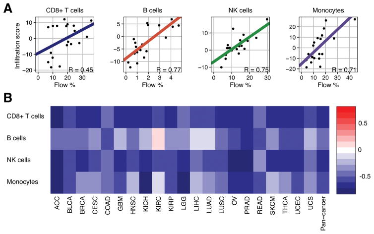
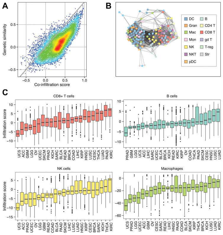
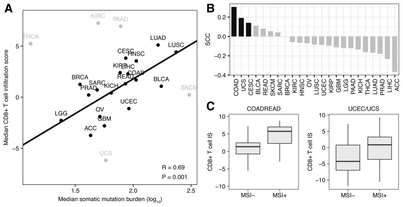
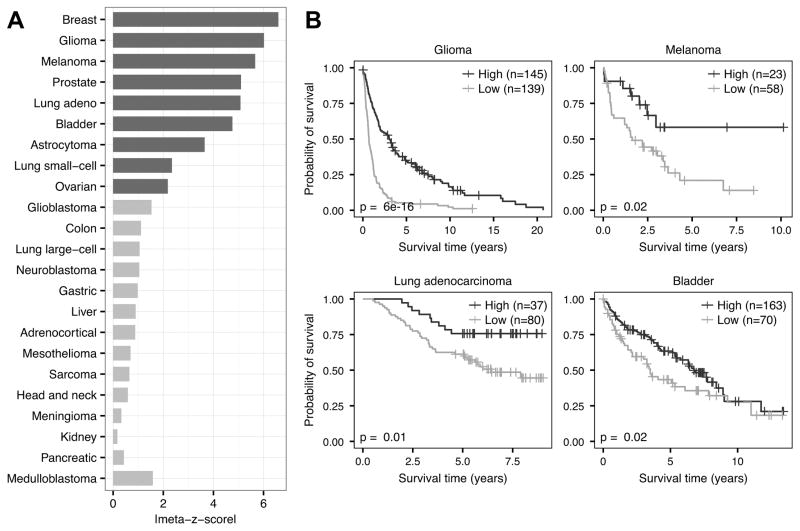
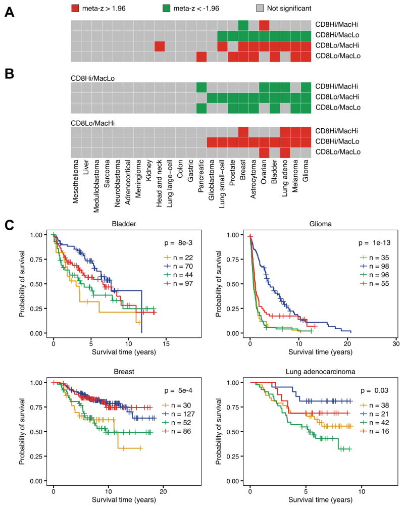
With the recent advent of immunotherapy, there is a critical need to understand immune cell interactions in the tumor microenvironment in both pan-cancer and tissue-specific contexts. Multidimensional datasets have enabled systematic approaches to dissect these interactions in large numbers of patients, furthering our understanding of the patient immune response to solid tumors. Using an integrated approach, we inferred the infiltration levels of distinct immune cell subsets in 23 tumor types from The Cancer Genome Atlas. From these quantities, we constructed a coinfiltration network, revealing interactions between cytolytic cells and myeloid cells in the tumor microenvironment. By integrating patient mutation data, we found that while mutation burden was associated with immune infiltration differences between distinct tumor types, additional factors likely explained differences between tumors originating from the same tissue. We concluded this analysis by examining the prognostic value of individual immune cell subsets as well as how coinfiltration of functionally discordant cell types associated with patient survival. In multiple tumor types, we found that the protective effect of CD8+ T cell infiltration was heavily modulated by coinfiltration of macrophages and other myeloid cell types, suggesting the involvement of myeloid-derived suppressor cells in tumor development. Our findings illustrate complex interactions between different immune cell types in the tumor microenvironment and indicate these interactions play meaningful roles in patient survival. These results demonstrate the importance of personalized immune response profiles when studying the factors underlying tumor immunogenicity and immunotherapy response. Cancer Res; 77(6); 1271-82. ©2017 AACR.


Conflict of interest statement

Disclosure of Potential Conflicts of Interests

The authors declare they have no competing interests.

Figures

Figure 1
Figure 1
Flow cytometry and tumor purity validation. a Scatterplot of flow cytometry and infiltration score for the four indicated immune cell subsets from 20 subjects. b Spearman correlations between infiltration scores from four indicated immune cell subsets and consensus purity estimates for 21 different cancer types. TCGA abbreviations for each cancer type are listed in Supplementary Table S1.
Figure 2
Figure 2
Immune cell co-infiltration analyses. a Scatterplot comparing the pairwise infiltration score Spearman correlation coefficients from all possible immune cell combinations (co-infiltration score) to pairwise expression Spearman correlation coefficients from all possible immune cell combinations (genetic similarity score). Gray line represents the trend if the genetic similarity scores were equal to their corresponding co-infiltration scores (y = x). Warmer colors represent higher point density while cooler colors lower point density. b Co-infiltration network representing correlation structure between each reference immune cell. Nodes represent reference immune cells. Edges represent co-infiltration scores > 0.45. Darker edges represent lower transcriptional similarity between reference cell types. c Boxplots comparing the distributions of immune infiltration in four indicated immune cell subsets. Each box spans quartiles with the lines representing the median correlation coefficient for each group. Whiskers represent absolute range excluding outliers. All outliers were included in the plot. TCGA abbreviations for each cancer type are listed in Supplementary Table S1.
Figure 3
Figure 3
Mutation burden and CD8+ T cell infiltration. A, Scatterplot of median somatic mutation number per tumor sample across 23 cancer types (log10 scale) compared with each tumor’s corresponding median CD8+ T cell infiltration score. Pearson correlation coefficient and least-squares regression line presented were calculated excluding the five outlier tumor types in gray. B, Individual Spearman correlation coefficients representing the associations between somatic mutation number and CD8+ T cell infiltration score in 23 different tumor types. Black bars represent statistically significant associations (P < 0.1). C, Box plots comparing the CD8+ T cell infiltration score (CD8+ T cell IS) in MSI− versus MSI+ samples. Left boxplot makes comparison in colorectal adenocarcinoma (COADREAD) samples. Right boxplot makes comparison in combined cohort of uterine corpus endometrial carcinoma (UCEC) and uterine carcinosarcoma (UCS) samples. Each box spans quartiles, with the lines representing the median correlation coefficient for each group. Whiskers represent absolute range excluding outliers. All outliers were included in the plot. TCGA abbreviations for each cancer type are listed in Supplementary Table S1.
Figure 4
Figure 4
Survival meta-analysis of CD8+ T cell infiltration. A, Meta-z-score absolute values indicating prognostic associations of CD8+ T cell infiltration in 23 different tumor types comprising 18,190 samples. Cancers were ranked by weighted meta-z-score. Dark gray bars indicate a weighted absolute meta-z-score >1.96, whereas light gray bars indicate a weighted meta-z-score whose absolute value is <1.96. B, Kaplan–Meier plots depicting the survival probability over time for samples with high (dark gray) and low (light gray) CD8+ T cell infiltration scores. Datasets tested include GSE16011 (glioma), GSE8401 (melanoma), GSE13213 (lung adenocarcinoma), and GSE5479 (bladder). For all Kaplan–Meier plots, samples were stratified into high and low groups based on their infiltration score distributions (thresholds available in Supplementary Figure S4). P values were calculated using the log-rank test. Vertical hash marks indicate censored data.
Figure 5
Figure 5
Effect of CD8+ T cell and macrophage infiltration on patient survival. a Weighted meta-z-scores from binary Cox proportional hazard models comparing the indicated class to the three remaining classes. b Weighted meta-z-scores from binary Cox proportional hazards models comparing CD8 T high/macrophage low (top) and CD8 T low/macrophage high (bottom) to the indicated classes. For all heatmaps, high/low status for each cell type was determined using median infiltration score for CD8+ T cells and macrophages. Red boxes indicate cancers where indicated class had significantly worse survival compared to other classes (meta-z > 1.96), green boxes indicate significantly improved survival compared to other classes (meta-z < -1.96), and gray boxes indicate no statistical association. c Kaplan-Meier plots depicting the survival distributions of all four classes: CD8+ T low/macrophage low (orange), CD8+ T high/macrophage low (blue), CD8+ T low/macrophage high (green), and CD8+ T high/macrophage high (red). Colors correspond to the classes as noted in a. Datasets tested include GSE5479 (bladder), GSE16011 (glioma), van de Vijver et al (breast), and GSE13213 (lung adenocarcinoma). For all Kaplan-Meier plots, samples were stratified into high and low groups based on their infiltration score distributions (thresholds available in Supplementary Figures S6 and S7). P-values were calculated using the log-rank test and indicate that at least one curve is significantly different from the rest. Vertical hash marks indicate censored data.

References

    1. Postow MA, Callahan MK, Wolchok JD. Immune Checkpoint Blockade in Cancer Therapy. J Clin Oncol. 2015;33:1974–82. - PMC - PubMed
    1. Maio M, Grob JJ, Aamdal S, Bondarenko I, Robert C, Thomas L, et al. Five-year survival rates for treatment-naive patients with advanced melanoma who received ipilimumab plus dacarbazine in a phase III trial. J Clin Oncol. 2015;33:1191–6. - PMC - PubMed
    1. Champiat S, Lambotte O, Barreau E, Belkhir R, Berdelou A, Carbonnel F, et al. Management of immune checkpoint blockade dysimmune toxicities: a collaborative position paper. Ann Oncol. 2016;27:559–74. - PubMed
    1. van Rooij N, van Buuren MM, Philips D, Velds A, Toebes M, Heemskerk B, et al. Tumor exome analysis reveals neoantigen-specific T-cell reactivity in an ipilimumab-responsive melanoma. J Clin Oncol. 2013;31:e439–42. - PMC - PubMed
    1. Tumeh PC, Harview CL, Yearley JH, Shintaku IP, Taylor EJ, Robert L, et al. PD-1 blockade induces responses by inhibiting adaptive immune resistance. Nature. 2014;515:568–71. - PMC - PubMed

Publication types

Substances