. 2017 Feb 14;114(7):E1118-E1127.
doi: 10.1073/pnas.1617705114. Epub 2017 Jan 26.

Design principles for robust vesiculation in clathrin-mediated endocytosis

Affiliations

Design principles for robust vesiculation in clathrin-mediated endocytosis

Julian E Hassinger et al. Proc Natl Acad Sci U S A. .

Abstract

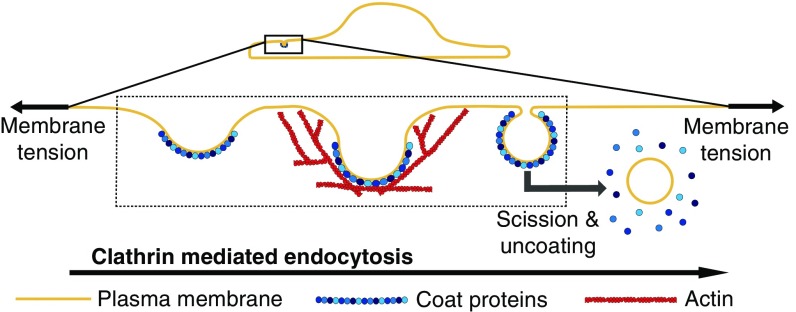
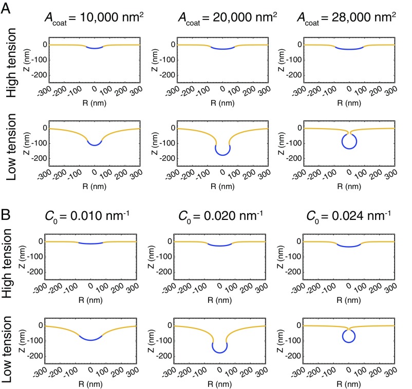
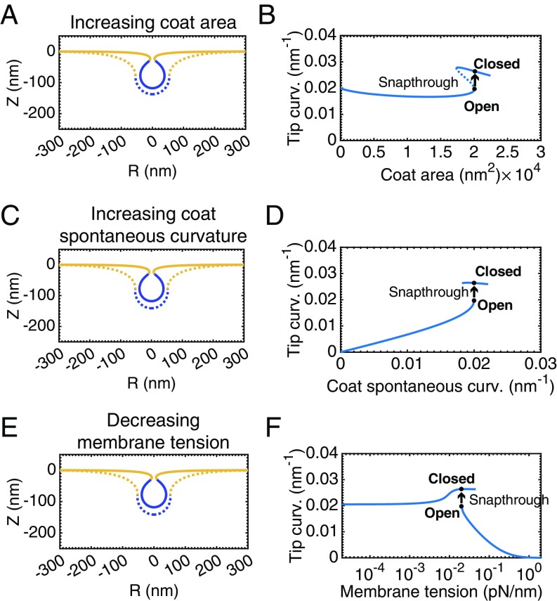
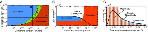
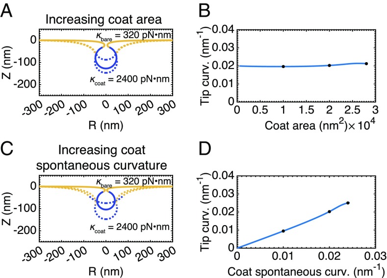
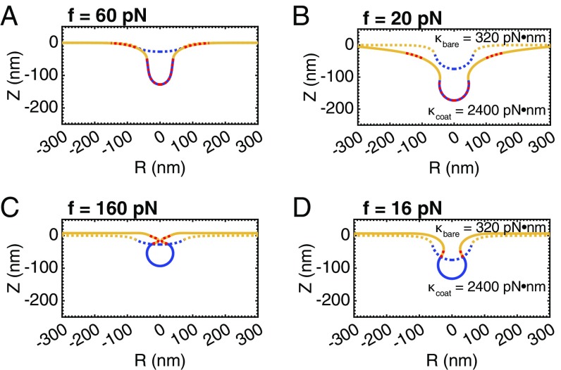
A critical step in cellular-trafficking pathways is the budding of membranes by protein coats, which recent experiments have demonstrated can be inhibited by elevated membrane tension. The robustness of processes like clathrin-mediated endocytosis (CME) across a diverse range of organisms and mechanical environments suggests that the protein machinery in this process has evolved to take advantage of some set of physical design principles to ensure robust vesiculation against opposing forces like membrane tension. Using a theoretical model for membrane mechanics and membrane protein interaction, we have systematically investigated the influence of membrane rigidity, curvature induced by the protein coat, area covered by the protein coat, membrane tension, and force from actin polymerization on bud formation. Under low tension, the membrane smoothly evolves from a flat to budded morphology as the coat area or spontaneous curvature increases, whereas the membrane remains essentially flat at high tensions. At intermediate, physiologically relevant, tensions, the membrane undergoes a "snap-through instability" in which small changes in the coat area, spontaneous curvature or membrane tension cause the membrane to "snap" from an open, U-shape to a closed bud. This instability can be smoothed out by increasing the bending rigidity of the coat, allowing for successful budding at higher membrane tensions. Additionally, applied force from actin polymerization can bypass the instability by inducing a smooth transition from an open to a closed bud. Finally, a combination of increased coat rigidity and force from actin polymerization enables robust vesiculation even at high membrane tensions.

Keywords: clathrin-mediated endocytosis; membrane modeling; membrane tension.


Conflict of interest statement

The authors declare no conflict of interest.

Figures

Fig. 1.
Fig. 1.
Schematic depiction of the main mechanical steps in CME. A multicomponent protein coat forms on the plasma membrane and causes the membrane to bend inward, forming a shallow pit. As the coat matures, the membrane becomes deeply invaginated to form an open, U-shaped pit before constricting to form a closed, Ω-shaped bud. The bud subsequently undergoes scission to form an internalized vesicle, and the coat is recycled. Actin polymerization is thought to provide a force, 𝐟, to facilitate these morphological changes, particularly at high membrane tensions (5). Our study is focused on understanding the impact of membrane tension on the morphological changes effected by the coat and actin polymerization, as indicated by the dashed box.
Fig. 2.
Fig. 2.
Membrane tension inhibits the ability of curvature generating coats to induce budding. (A) Profile views of membrane morphologies generated by simulations in which the area of a curvature-generating coat progressively increases, covering more of the bare membrane. The curvature-generating capability, or spontaneous curvature, of the coat is set at C0= 0.02nm1, corresponding to a preferred radius of curvature of 50nm (12). (A, Upper) High membrane tension, λ0= 0.2pN/nm. The membrane remains nearly flat as the area of the coat increases. (A, Lower) Low membrane tension, λ0= 0.002pN/nm. Addition of coat produces a smooth evolution from a flat membrane to a closed bud. (B) Membrane profiles for simulations with a constant coat area in which the spontaneous curvature of the coat progressively increases. The area of the coat is Acoat= 20,106nm2. (B, Upper) High membrane tension, λ0= 0.2pN/nm. The membrane remains nearly flat with increasing spontaneous curvature. (B, Lower) Low membrane tension, λ0= 0.002pN/nm. Increasing the spontaneous curvature of the coat induces a smooth evolution from a flat membrane to a closed bud.
Fig. 3.
Fig. 3.
A snap-through instability exists at intermediate, physiologically relevant (54), membrane tensions, λ0= 0.02pN/nm. (A) Membrane profiles showing bud morphology before (dashed line, Acoat= 20,065nm2) and after (solid line, Acoat= 20,105nm2) addition of a small amount of area to the coat, C0= 0.02nm1. (B) Mean curvature at the tip of the bud as a function of the coat area. There are two stable branches of solutions of the equilibrium membrane shape equations. The lower branch consists of open, U-shaped buds, whereas the upper branch consists of closed, Ω-shaped buds. The dashed portion of the curve indicates “unstable” solutions that are not accessible by simply increasing and decreasing the area of the coat. The marked positions on the curve denote the membrane profiles shown in A. The transition between these two shapes is a snap-through instability, in which the bud snaps closed upon a small addition to area of the coat. (C) Bud morphologies before (dashed line) and after (solid line) a snap-through instability with increasing spontaneous curvature, Acoat=20,106nm2, C0= 0.02nm2. (D) Mean curvature at the tip of the bud as a function of the spontaneous curvature of the coat. (E) Bud morphology before (dashed line) and after (solid line) a snap-through instability with decreasing membrane tension, Acoat= 20,106nm2, C0= 0.02nm2, λ0= 0.02pN/nm. (F) Mean curvature at the tip of the bud as a function of the membrane tension.
Fig. 4.
Fig. 4.
Bud morphology depends on bending rigidity, membrane tension, spontaneous curvature, and coat area. (A) Coat spontaneous curvature (C0) vs. membrane tension (λ0) phase diagram. The regions of the diagram are color coded according to the final shape of the membrane for coat “growing” simulations performed with the specified values for edge membrane tension and coat spontaneous curvature. Blue denotes closed, Ω-buds; red denotes open, U-shaped pits; and green are situations in which closed buds are obtained via a snap-through transition. The snap-through solutions cluster about the dashed line, Ves=1, which separates the high and low membrane tension regimes (for details, see The Instability Exists over a Range of Membrane Tensions, Coat Areas, and Spontaneous Curvatures). The lines labeled B and C, respectively, indicate the phase diagrams at right. (B) Coat area vs. membrane tension phase diagram, C0=0.02nm1. Blue denotes closed buds, red denotes open buds, and green denotes parameters that have both open and closed bud solutions. The dashed line, Ves=1, marks the transition from low to high membrane tension. The solid line represents the theoretical area of a sphere that minimizes the Helfrich energy at the specified membrane tension (SI Appendix, 3. Radius of a Vesicle from Energy Minimization). (C) Coat area vs. spontaneous curvature phase diagram, λ0=0.02pN/nm. The dashed line, Ves=1, marks the transition between spontaneous curvatures that are capable and incapable of overcoming the membrane tension to form a closed bud. The solid line represents the theoretical area of a sphere that minimizes the Helfrich energy at the specified spontaneous curvature (SI Appendix, 3. Radius of a Vesicle from Energy Minimization).
Fig. 5.
Fig. 5.
The snap-through instability at physiological tension, λ0=0.02pN/nm, is abolished when the bending rigidity of the coat is increased relative to the bare membrane, κbare= 320pNnm, κcoat=2400pNnm. (A) Membrane profiles showing a smooth progression of bud morphologies as the area of the coat is increased (Acoat= 10,000nm2, 20,000nm2, 28,000nm2), C0= 0.02nm1. (B) Mean curvature at the bud tip as a function of the area of the coat. The marked positions denote the membrane profiles shown in A. There is now only a single branch of solutions (compared with Fig. 3B), indicating a smooth evolution from a flat membrane to a closed bud. (C) Membrane profiles showing a smooth progression of bud morphologies as spontaneous curvature of the coat is increased (C0= 0.01nm1, 0.02nm1, 0.024nm1), Acoat= 20,106nm2. (D) Mean curvature at the bud tip as a function of the spontaneous curvature of the coat showing a single branch of solutions (compare with Fig. 3D).
Fig. 6.
Fig. 6.
A force from actin assembly can mediate the transition from a U- to Ω-shaped bud, avoiding the instability at intermediate membrane tension, λ0= 0.02pN/nm. Two orientations of the actin force were chosen based on experimental evidence from yeast (31) and mammalian (45) cells. (A) Schematic depicting actin polymerization in a ring at the base of the pit with the network attached to the coat, causing a net inward force on the bud. (B) At constant coat area, Acoat= 17,593nm2, and spontaneous curvature, C0= 0.02nm1, a force (red dash) adjacent to the coat drives the shape transition from a U-shaped (dashed line) to Ω-shaped bud (solid line). The force intensity was homogeneously applied to the entire coat, and the force intensity at the base of the pit was set such that the total force on the membrane integrates to zero. The final applied inward force on the bud was 𝐟= 15pN, well within the capability of a polymerizing actin network (60). (C) Schematic depicting actin assembly in a collar at the base, directly providing a constricting force (45). (D) A constricting force (red dash) localized to the coat drives the shape transition from a U-shaped (dashed line) to Ω-shaped bud (solid line), Acoat= 17,593nm2, C0= 0.02nm1. The force intensity was homogeneously applied perpendicular to the membrane to an area of 5,027nm2 immediately adjacent to the coated region. The final applied force on the membrane was 𝐟< 1pN.
Fig. 7.
Fig. 7.
A combination of increased coat rigidity and force from actin polymerization ensures robust vesiculation, even at high membrane tension, λ0= 0.2pN/nm, C0= 0.02nm1. (A) Application of the inward directed actin force (as in Fig. 6A) induces tubulation, but not vesiculation, at high tension. (B) Increasing the stiffness of the coat alone is insufficient to overcome high membrane tension (dashed line). However, increasing the coat stiffness enables the applied force to induce vesiculation and decreases the magnitude of the force required by a factor of 3. (C) Application of the constricting actin force (as in Fig. 6C) is sufficient to induce vesiculation, even at high tension. The magnitude of the applied force required is likely unrealistically high in a biologically relevant setting. (D) Increasing the coat stiffness decreases the force required to induce vesiculation by an order of magnitude.
Fig. 8.
Fig. 8.
Design principles for robust vesiculation. The rigidity of the plasma membrane, as well as the membrane tension, resists budding by curvature-generating coats. In the low tension regime, as defined by the vesiculation number, increasing the coat area or spontaneous curvature is sufficient to induce a smooth evolution from a flat membrane to a closed bud. A combination of increased coat rigidity and force from actin polymerization is necessary to ensure robust vesiculation in the high membrane-tension regime.

Similar articles

Cited by

References

    1. Johannes L, Wunder C, Bassereau P. Bending “on the rocks” cocktail of biophysical modules to build endocytic pathways. Cold Spring Harb Perspect Biol. 2014;6(1):a016741. - PMC - PubMed
    1. Taylor MJ, Perrais D, Merrifield CJ. A high precision survey of the molecular dynamics of mammalian clathrin-mediated endocytosis. PLoS Biol. 2011;9(3):e1000604. - PMC - PubMed
    1. Liu J, Sun Y, Drubin DG, Oster GF. The mechnochemistry of endocytosis. PLoS Biol. 2009;7(9):e1000204. - PMC - PubMed
    1. Liu J, Sun Y, Oster GF, Drubin DG. Mechanochemical crosstalk during endocytic vesicle formation. Curr Opin Cell Biol. 2010;22(1):36–43. - PMC - PubMed
    1. Boulant S, Kural C, Zeeh JC, Ubelmann F, Kirchhausen T. Actin dynamics counteract membrane tension during clathrin-mediated endocytosis. Nat Cell Biol. 2011;13(9):1124–1131. - PMC - PubMed

Publication types