. 2017 Mar;58(3):563-577.
doi: 10.1194/jlr.M073510. Epub 2017 Jan 26.

Lipids regulate the hydrolysis of membrane bound glucosylceramide by lysosomal β-glucocerebrosidase

Affiliations

Lipids regulate the hydrolysis of membrane bound glucosylceramide by lysosomal β-glucocerebrosidase

Misbaudeen Abdul-Hammed et al. J Lipid Res. 2017 Mar.

Abstract

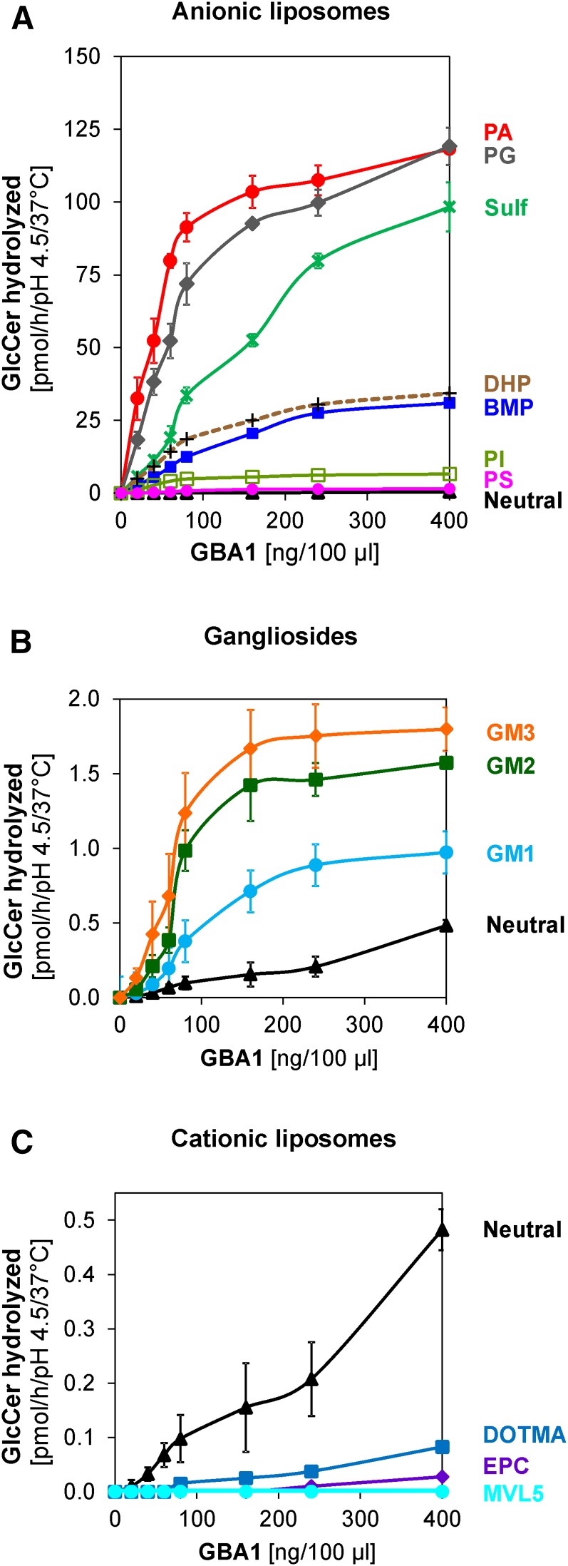
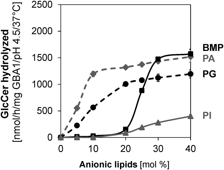
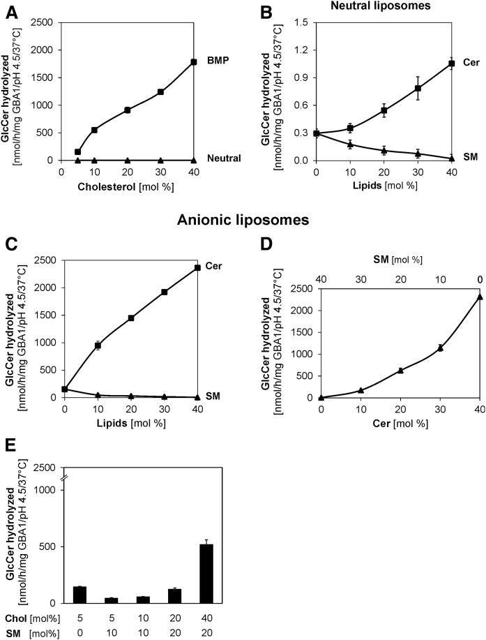
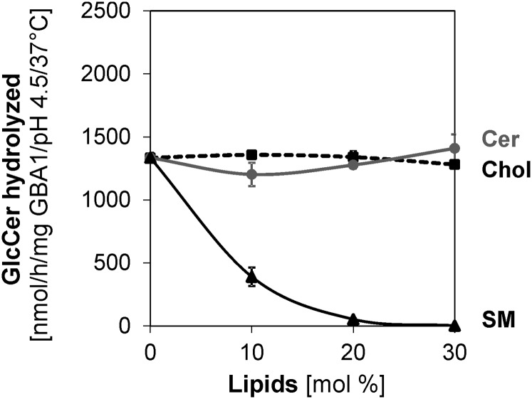
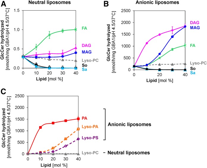
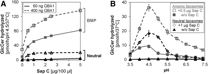
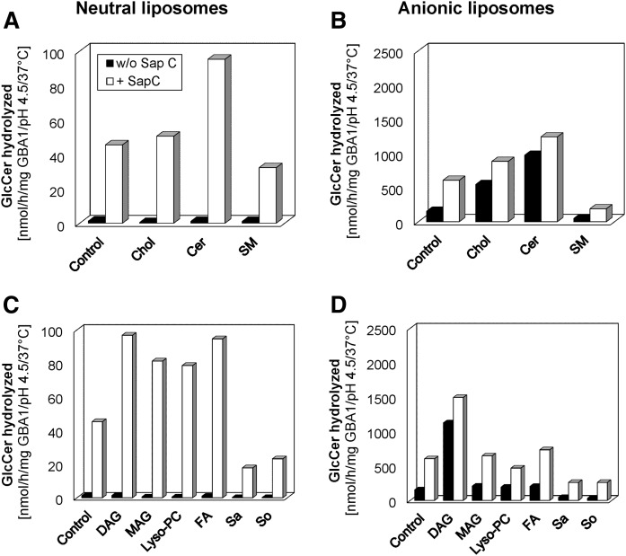
Glucosylceramide (GlcCer) is the primary storage lipid in the lysosomes of Gaucher patients and a secondary one in Niemann-Pick disease types A, B, and C. The regulatory roles of lipids on the hydrolysis of membrane bound GlcCer by lysosomal β-glucocerebrosidase (GBA1) was probed using a detergent-free liposomal assay. The degradation rarely occurs at uncharged liposomal surfaces in the absence of saposin (Sap) C. However, anionic lipids stimulate GlcCer hydrolysis at low pH by up to 1,000-fold depending on the nature and position of the negative charges in their head groups while cationic lipids inhibit the degradation, thus showing the importance of electrostatic interactions between the polycationic GBA1 and the negatively charged vesicle surfaces at low pH. Ceramide, fatty acids, monoacylglycerol, and diacylglycerol also stimulate GlcCer hydrolysis while SM, sphingosine, and sphinganine play strong inhibitory roles, thereby explaining the secondary storage of GlcCer in Niemann-Pick diseases. Surprisingly, cholesterol stimulates GlcCer degradation in the presence of bis(monoacylglycero)phosphate (BMP). Sap C strongly stimulates GlcCer hydrolysis even in the absence of BMP and the regulatory roles of the intraendolysosomal lipids on its activity is discussed. Our data suggest that these strong modifiers of GlcCer hydrolysis affect the genotype-phenotype correlation in several cases of Gaucher patients independent of the types.

Keywords: Gaucher disease; acylglycerols; anionic phospholipids; cationic lipids; cholesterol; electrostatic interaction; fatty acids; lysophospholipids; sphingomyelin; sphingosine.


Figures

Fig. 1.
Fig. 1.
Assay optimization for the hydrolysis of membrane bound GlcCer by GBA1. Neutral liposomes contain 5 mol% cholesterol, 1 mol% [14C]GlcCer, and 94 mol% DOPC. Anionic liposomes contain 20 mol% anionic lipid (BMP or PA), 5 mol% cholesterol, 1 mol% [14C]GlcCer, and 74 mol% DOPC. The enzyme concentration is 60 or 400 ng GBA1/100 μl assay in anionic and neutral liposomes, respectively, and the experiments were carried out at pH 4.5 under 30 min incubation and at 37°C (except as otherwise stated). GlcCer hydrolysis is enzyme dose dependent (A) and time dependent (B) in neutral (black), BMP-containing (blue), and PA-containing liposomes (red). GBA1 activity is pH dependent in PA-containing (C) and BMP-containing (D) liposomes. High ionic strength inhibits the hydrolysis of GlcCer by GBA1 even in the presence 20 mol% PA (E) or 20 mol% BMP (F), using 20 mM (red) and 50 mM (blue) citrate buffers (pH 4.5) containing 0–300 mM NaCl. Mean ± SEM (n = 4–6).
Fig. 2.
Fig. 2.
Regulatory roles of anionic and cationic membrane lipids on the hydrolysis of membrane bound GlcCer by GBA1. A: GlcCer digestion is strongly stimulated by PA (red), PG (gray), and sulfatide (green) and moderately stimulated by DHP (brown), BMP (blue), and PI (light green), but only weakly stimulated by PS (pink). B: Only weak stimulations are observed with gangliosides; GM1 (light blue), GM2 (green), and GM3 (orange) as well as in neutral (black) liposomes, while cationic lipids, such as EPC (purple), DOTMA (blue), and MVL5 (light blue), inhibit the GlcCer hydrolysis (C). Mean ± SEM (n = 4–6).
Fig. 3.
Fig. 3.
Anionic lipids stimulate membrane bound GlcCer hydrolysis by GBA1 in a concentration-dependent manner. Liposomes containing 0–40 mol% anionic lipids (PI, PG, PA, or BMP) were used. The experiments were carried out using 60 ng GBA1 per assay under 30 min incubation and at 37°C. Mean ± SEM (n = 4–6).
Fig. 4.
Fig. 4.
Cholesterol and Cer strongly stimulate membrane bound GlcCer hydrolysis by GBA1, while SM plays a strong inhibitory role. Cholesterol strongly stimulates GlcCer hydrolysis in the presence of BMP (A). Liposomes contain 5–40 mol% cholesterol, 1 mol% [14C]GlcCer (Neutral), and, in addition, 20 mol% BMP. Cer stimulates GlcCer hydrolysis, while SM displays an inhibitory role in the neutral liposomes (B) and in the presence of BMP (C). Liposomes contain 5 mol% cholesterol, 1 mol% [14C]GlcCer (Neutral), and, in addition, 20 mol% BMP with various concentrations of Cer and SM. GlcCer hydrolysis is strongly stimulated as SM is gradually replaced with Cer (D) in the presence of BMP with the total SM and Cer contents being 40 mol% at any time. Cholesterol plays stimulatory roles even in the presence of SM (E) in BMP-containing liposomes. The experiments were carried out using 10,000 and 60 ng GBA1 per assay for neutral and BMP-containing liposomes, respectively, at pH 4.5, under 30 min incubation and at 37°C. Mean ± SEM (n = 4–6).
Fig. 5.
Fig. 5.
In the presence of PA as the anionic lipid, cholesterol (Chol), and Cer regulatory effects on membrane bound GlcCer hydrolysis by GBA1 are insignificant, while SM plays a strong inhibitory role. Liposomes contain 5 mol% cholesterol, 1 mol% [14C]GlcCer, 20 mol% PA, and 0–30 mol% cholesterol, Cer, or SM (as indicated) with DOPC being the host lipid. The experiments were carried out using 60 ng GBA1 per assay under 30 min incubation and at 37°C. Mean ± SEM (n = 4–6).
Fig. 6.
Fig. 6.
Stimulatory and inhibitory roles of various lysosomal lipid degradation products on membrane bound GlcCer hydrolysis by GBA1. Stearic acid, FA (green), DAG (pink) and MAG (blue) stimulate GlcCer hydrolysis; sphingosine (So) (black) and sphinganine (Sa) (light blue) strongly inhibit GlcCer hydrolysis, while lyso-PC (gray) plays different roles in neutral liposomes (A) and in the presence of BMP (B). In the absence of BMP (neutral liposomes), lyso-phospholipids carrying negatively charged head groups [lyso-PA (orange) and lyso-PS (purple), as indicated] strongly stimulate GlcCer hydrolysis more highly than the neutral lyso-PC, but to a lesser extent compared with PA (red) (C). The experiments were carried out using 10,000 and 60 ng GBA1/100 μl assay for neutral and anionic liposomes, respectively, at pH 4.5, under 30 min incubation and at 37°C. Mean ± SEM (n = 4–6).
Fig. 7.
Fig. 7.
Sap C facilitates the hydrolysis of membrane bound GlcCer by GBA1 even in the absence of anionic phospholipid (BMP). GlcCer hydrolysis is Sap C dose dependent (A) and the Sap C-induced GlcCer hydrolysis by GBA1 is significant even in the absence of BMP, but at higher GBA1 concentration. However, the presence of BMP tremendously enhances the Sap C activity. The pH profile of GlcCer hydrolysis becomes narrower in the presence of both Sap C and anionic lipid, BMP (B). The pH optimum for all experiments remains 4.5 in the presence or absence of Sap C, with the narrowest pH profile obtained during the combined presence of both Sap C and BMP. The experiments were carried out using 400 and 60 ng GBA1 per assay for neutral and anionic liposomes (containing 20 mol% BMP), respectively, at pH 4.5, under 30 min incubation and at 37°C. Mean – SEM (n = 4–6).
Fig. 8.
Fig. 8.
Stimulatory and inhibitory roles of various lysosomal lipid degradation products on Sap C-induced hydrolysis of membrane bound GlcCer by GBA1. GlcCer hydrolysis is insignificant in the absence of both Sap C and BMP. Sap C-induced GlcCer hydrolysis by GBA1 is enhanced strongly by Cer and slightly by cholesterol (Chol), but inhibited by SM in neutral liposomes (A) and in the presence of BMP (B), respectively. Also, the Sap C-induced GlcCer hydrolysis by GBA1 is strongly enhanced by DAG, MAG, stearic acid, and lyso-PC, but strongly inhibited by sphingosine (So) and sphinganine (Sa) in neutral liposomes (C). In the presence of BMP (D), it is enhanced strongly by DAG and slightly by MAG and stearic acid, but strongly inhibited by sphingosine and sphinganine. The effects of lyso-PC depend on the liposome types. The experiments were carried out using 10 mol% each of the stimulating or inhibiting lipids and 400 or 60 ng GBA1 per assay with 1 or 0.5 μg Sap C for neutral and BMP-containing liposomes, respectively, under 30 min incubation and at 37°C. SEM was less than 10% (n = 4–6).

References

    1. Schulze H., Kolter T., and Sandhoff K.. 2009. Principles of lysosomal membrane degradation: Cellular topology and biochemistry of lysosomal lipid degradation. Biochim. Biophys. Acta. 1793: 674–683. - PubMed
    1. Brady R. O., Kanfer J. N., and Shapiro D.. 1965. Metabolism of glucocerebrosides. II. Evidence of an enzymatic deficiency in Gaucher’s disease. Biochem. Biophys. Res. Commun. 18: 221–225. - PubMed
    1. Eckford P. D., and Sharom F. J.. 2005. The reconstituted P-glycoprotein multidrug transporter is a flippase for glucosylceramide and other simple glycosphingolipids. Biochem. J. 389: 517–526. - PMC - PubMed
    1. Sasaki T. 1990. Glycolipid transfer protein and intracellular traffic of glucosylceramide. Experientia. 46: 611–616. - PubMed
    1. Möbius W., Herzog V., Sandhoff K., and Schwarzmann G.. 1999. Intracellular distribution of a biotin-labeled ganglioside, GM1, by immunoelectron microscopy after endocytosis in fibroblasts. J. Histochem. Cytochem. 47: 1005–1014. - PubMed

Publication types

MeSH terms