Review
. 2017:2017:8385961.
doi: 10.1155/2017/8385961. Epub 2017 Jan 3.

Sterile Neuroinflammation and Strategies for Therapeutic Intervention

Affiliations
Review

Sterile Neuroinflammation and Strategies for Therapeutic Intervention

Manoj Banjara et al. Int J Inflam. 2017.

Abstract

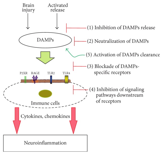
Sterile neuroinflammation is essential for the proper brain development and tissue repair. However, uncontrolled neuroinflammation plays a major role in the pathogenesis of various disease processes. The endogenous intracellular molecules so called damage-associated molecular patterns or alarmins or damage signals that are released by activated or necrotic cells are thought to play a crucial role in initiating an immune response. Sterile inflammatory response that occurs in Alzheimer's disease (AD), Parkinson's disease (PD), stroke, hemorrhage, epilepsy, or traumatic brain injury (TBI) creates a vicious cycle of unrestrained inflammation, driving progressive neurodegeneration. Neuroinflammation is a key mechanism in the progression (e.g., AD and PD) or secondary injury development (e.g., stroke, hemorrhage, stress, and TBI) of multiple brain conditions. Hence, it provides an opportunity for the therapeutic intervention to prevent progressive tissue damage and loss of function. The key for developing anti-neuroinflammatory treatment is to minimize the detrimental and neurotoxic effects of inflammation while promoting the beneficial and neurotropic effects, thereby creating ideal conditions for regeneration and repair. This review outlines how inflammation is involved in the pathogenesis of major nonpathogenic neuroinflammatory conditions and discusses the complex response of glial cells to damage signals. In addition, emerging experimental anti-neuroinflammatory drug treatment strategies are discussed.


Conflict of interest statement

The authors declare that they have no competing interests.

Figures

Figure 1
Figure 1
Scheme of early innate response to brain injury. Damage signals or DAMPs primarily released from the injured parenchymal cells are sensed by immune effector cells such as microglia, astrocytes, and macrophages. The triggered innate immune response (e.g., proinflammatory cytokines, chemokines, reactive oxygen species, excitotoxins, histamine, and prostaglandins) has detrimental influences on the neurons, oligodendroglial precursors, and vascular endothelial cells. The increased BBB permeability contributes the migration of peripheral immune cells (e.g., neutrophils, mast cells, and macrophages) to the sites of tissue damage.
Figure 2
Figure 2
Response of microglia and astrocytes to the brain injury. DAMPs can signal PRRs expressed in astrocytes and microglia, promoting their activation. Depending on the injury site, severity of brain injury, surrounding environment, and signaling strength, astrocytes and microglia may respond to remove stimulants or to secrete inflammatory mediators. Typically, beneficial activation (M2-like microglia and radial-glia-like astrocytes) is associated with the elevated release of neurotrophic factors, anti-inflammatory cytokines (e.g., IL-4 and IL-10), and enzymes (e.g., arginase 1 and insulin-degrading enzymes) that enhance phagocytic activity. Conversely, detrimental activation of astrocytes and microglia is associated with the elevated and sustained expression of inducible nitric oxide synthase, reactive oxygen species, proinflammatory mediators (e.g., IL-1α/β, IL-6, and TNF), and decreased secretion of neurotrophic factors. These divergent responses may determine whether microglia and astrocytes lead to clear tissue debris or promote chronic neuroinflammation.
Figure 3
Figure 3
Propagation of “damage signals.” Harmful stimuli in the brain (e.g., brain injury and excessive neurodegeneration) generate endogenous DAMPs that induce the release of inflammatory mediators by activating PRRs. In turn, these molecules upregulate their own expression, directly activate the release of DAMPs, and trigger further tissue damage leading to increasing DAMPs level. Hence, a sustained aggressive cycle may result in chronic neuroinflammation. However, a controlled release of DAMPs has beneficial roles in immunity and tissue repair process.
Figure 4
Figure 4
Mechanisms of glial cell activation in response to damage signals. Sterile neuroinflammatory conditions are characterized by the accumulation of misfolded and aggregated proteins in the brain. These DAMPs are released from different subcellular components of the damaged neurons, which trigger respective PRRs leading to downstream activation of proinflammatory cascades and enhancing effects of initial inflammatory insult. Activation of PRRs, primarily TLR2, TLR4, TLR9, and RAGE, converge largely into NF-κB activation, promoting cell death and/or contributing to neuroinflammatory/neurodegenerative mechanisms. These pathways, including P2XR, jointly work with multiprotein inflammasome complex (NLRs) that assists the generation of mature cytokines from proforms via the activation of caspase-1. TLR, toll-like receptor; RAGE, receptor for advanced glycation end products; NLR, nod-like receptor; P2XR, ATP-gated purinergic P2 receptors; MyD88, myeloid differentiation primary response gene (88); MAL, MyD88-adapter-like; TRIF, TIR-domain-containing adapter-inducing interferon-β; TRAM, TRIF-related adaptor molecule; MAPK, mitogen-activated protein kinase; NF-κB, nuclear factor-kappa B; IL, interleukin.
Figure 5
Figure 5
Drug treatment strategies for DAMPs-induced neuroinflammation. Preclinical studies have identified a number of multipotential drug targets that attenuates neuroinflammation triggered by DAMPs released after brain injury or excessive neurodegeneration.

References

    1. Fernandes-Alnemri T., Yu J.-W., Datta P., Wu J., Alnemri E. S. AIM2 activates the inflammasome and cell death in response to cytoplasmic DNA. Nature. 2009;458(7237):509–513. doi: 10.1038/nature07710. - DOI - PMC - PubMed
    1. Lyman M., Lloyd D. G., Ji X., Vizcaychipi M. P., Ma D. Neuroinflammation: the role and consequences. Neuroscience Research. 2014;79(1):1–12. doi: 10.1016/j.neures.2013.10.004. - DOI - PubMed
    1. de Vries H. E., Blom-Roosemalen M. C., van Oosten M., et al. The influence of cytokines on the integrity of the blood-brain barrier in vitro. Journal of Neuroimmunology. 1996;64(1):37–43. doi: 10.1016/0165-5728(95)00148-4. - DOI - PubMed
    1. Kitazawa M., Oddo S., Yamasaki T. R., Green K. N., LaFerla F. M. Lipopolysaccharide-induced inflammation exacerbates tau pathology by a cyclin-dependent kinase 5-mediated pathway in a transgenic model of Alzheimer's disease. The Journal of Neuroscience. 2005;25(39):8843–8853. doi: 10.1523/jneurosci.2868-05.2005. - DOI - PMC - PubMed
    1. Heneka M. T., Kummer M. P., Latz E. Innate immune activation in neurodegenerative disease. Nature Reviews Immunology. 2014;14(7):463–477. doi: 10.1038/nri3705. - DOI - PubMed