Review
. 2016:2016:5482087.
doi: 10.1155/2016/5482087. Epub 2016 Dec 29.

Mucosal Vaccine Development Based on Liposome Technology

Affiliations
Review

Mucosal Vaccine Development Based on Liposome Technology

Valentina Bernasconi et al. J Immunol Res. 2016.

Abstract

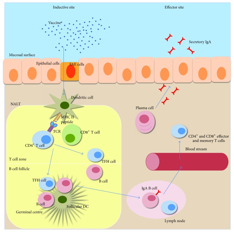
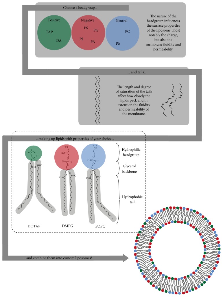
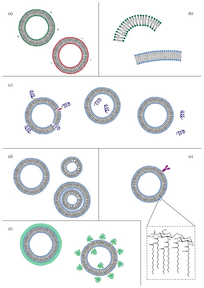
Immune protection against infectious diseases is most effective if located at the portal of entry of the pathogen. Hence, there is an increasing demand for vaccine formulations that can induce strong protective immunity following oral, respiratory, or genital tract administration. At present, only few mucosal vaccines are found on the market, but recent technological advancements and a better understanding of the principles that govern priming of mucosal immune responses have contributed to a more optimistic view on the future of mucosal vaccines. Compared to live attenuated vaccines, subcomponent vaccines, most often protein-based, are considered safer, more stable, and less complicated to manufacture, but they require the addition of nontoxic and clinically safe adjuvants to be effective. In addition, another limiting factor is the large antigen dose that usually is required for mucosal vaccines. Therefore, the combination of mucosal adjuvants with the recent progress in nanoparticle technology provides an attractive solution to these problems. In particular, the liposome technology is ideal for combining protein antigen and adjuvant into an effective mucosal vaccine. Here, we describe and discuss recent progress in nanoparticle formulations using various types of liposomes that convey strong promise for the successful development of the next generation of mucosal vaccines.


Conflict of interest statement

The authors have no conflict of interests.

Figures

Figure 1
Figure 1
Principles for induction of mucosal immune responses after intranasal vaccination. The respiratory mucosal immune system consists of clusters of lymphoid cells beneath the mucosal epithelium, hosting both innate and adaptive immune cells [29]. There is a clear distinction between inductive and effector sites and these are also physically separated. Inductive sites are organized lymphoid tissues where antigen is taken up by DCs and other APCs. The effector sites, on the other hand, are tissues that provide protection against infection where specific antibodies and CD4+ and CD8+ effector and memory T cells reside [30]. The main inductive sites for mucosal immune responses after intranasal vaccination are known as nasopharynx-associated lymphoid tissue (NALT), which harbors B cell follicles and T cell zones in well demarked microanatomical areas [31]. Antigens are taken up by DCs that get access to the luminal content either through direct uptake through the epithelium or via the follicle associated epithelium (FAE) that overlay the NALT. After antigen uptake, the immature DCs undergo maturation and subsequently leave the mucosal tissue for the draining lymph nodes, alternatively, if already in the NALT, the DCs will directly prime naive CD4+ or CD8+ T cells. Activated CD4+ T cells differentiate into various subsets: T helper 1 (Th1), Th2, or Th17 cells, regulatory T cells (Tregs), or follicular helper T cells (TFH). The latter are critically needed for the expansion and differentiation of the activated B cells in the germinal center (GC), which is formed in the B cell follicle in the lymph node after vaccination. TFH cells are involved in the development of long-lived plasma cells and memory B cells in the GC.
Figure 2
Figure 2
Generation of customized liposomes. Lipids are polar molecules consisting of a hydrophilic headgroup and hydrophobic fatty acid tails. Examples of positively charged headgroups are trimethylammonium propane (TAP) and dioctadecyl ammonium bromide (DA), while negatively charged headgroups are phosphatidylserine (PS), phosphatidylinositol (PI), phosphatidylglycerol (PG), or phosphatidic acid (PA), and finally neutral headgroups are phosphatidylcholine (PC) or phosphatidylethanolamine (PE). A headgroup can be combined with tails of different nature to create lipids with the desired properties; the examples shown are the lipid 1,2-dioleoyl-3-trimethylammonium propane (DOTAP) and the phospholipids dimyristoylphosphatidylglycerol (DMPG) and 1-palmitoyl-2-oleoyl-sn-glycero-3-phosphocholine (POPC). Different lipids can then be combined into liposomes with different functional features, which provide the basis for this highly diverse and versatile technology that is so excellently suited for vaccine development.
Figure 3
Figure 3
Various properties of liposome-based vaccines. (a) The effect of altered surface charge on liposome function has been extensively examined [32, 33]. (b) The lipid composition is critically influencing the immune response [34]. (c) Also the localization of the antigen on or inside the liposome plays an important role in shaping the immune response to the vaccine. There are several modes of antigen association to liposomes. Firstly, antigens may be encapsulated in the aqueous core or they could be linked to the surface via covalent attachment. Alternatively, a hydrophobic anchor can be used to attach the antigen to the surface via adsorption or through electrostatic interactions with lipids of opposite charge. For proteins with a hydrophobic region one may even successfully insert these in the liposome membrane. The liposome may also be used as an immunoenhancer simply by admixing the antigen and the liposomes. (d) Only few studies have addressed the impact of size or lamellarity [35, 36]. (e) Modifications of liposomes to increase their immunoenhancing effect can be done through attaching PAMPs, such as lipid A (LPS), or through specific targeting strategies using cell-specific antibodies (anti-CD103 or -DEC205) [16, 37]. (f) Other modifications, including addition of poly(ethylene glycol) (PEG) or different polymer coatings that increase the liposome penetration of the mucosal barrier or to increase liposome resistance in biological fluids, have also been developed [38].

References

    1. Kurono Y., Yamamoto M., Fujihashi K., et al. Nasal immunization induces Haemophilus influenzae-specific Th1 and Th2 responses with mucosal IgA and systemic IgG antibodies for protective immunity. Journal of Infectious Diseases. 1999;180(1):122–132. doi: 10.1086/314827. - DOI - PubMed
    1. Ryan E. J., Daly L. M., Mills K. H. G. Immunomodulators and delivery systems for vaccination by mucosal routes. Trends in Biotechnology. 2001;19(8):293–304. doi: 10.1016/S0167-7799(01)01670-5. - DOI - PubMed
    1. Levine M. M. Can needle-free administration of vaccines become the norm in global immunization? Nature Medicine. 2003;9(1):99–103. doi: 10.1038/nm0103-99. - DOI - PubMed
    1. Lycke N. Recent progress in mucosal vaccine development: potential and limitations. Nature Reviews Immunology. 2012;12(8):592–605. doi: 10.1038/nri3251. - DOI - PubMed
    1. Chadwick S., Kriegel C., Amiji M. Nanotechnology solutions for mucosal immunization. Advanced Drug Delivery Reviews. 2010;62(4-5):394–407. doi: 10.1016/j.addr.2009.11.012. - DOI - PubMed

LinkOut - more resources