

MicroRNA-9 inhibits the gastric cancer cell proliferation by targeting TNFAIP8
- PMID: 28127811
- PMCID: PMC6529077
- DOI: 10.1111/cpr.12331
MicroRNA-9 inhibits the gastric cancer cell proliferation by targeting TNFAIP8
Abstract
Background and objectives: MicroRNA-9 is frequently dysregulated in many human carcinoma types, including gastric cancer (GC). Previous studies demonstrated that the expression of TNFAIP8 in GC is correlated with tumour occurrence, development, invasion, metastasis and prognosis. However, till now, the relationship between MicroRNA-9 and TNFAIP8 in GC has not been reported.
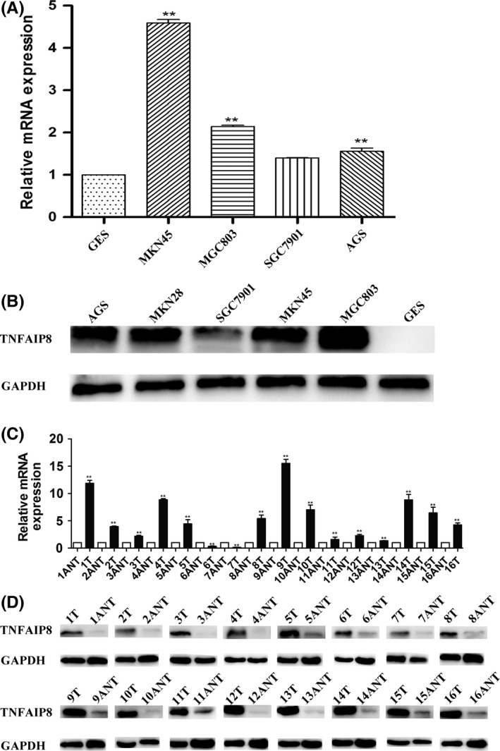
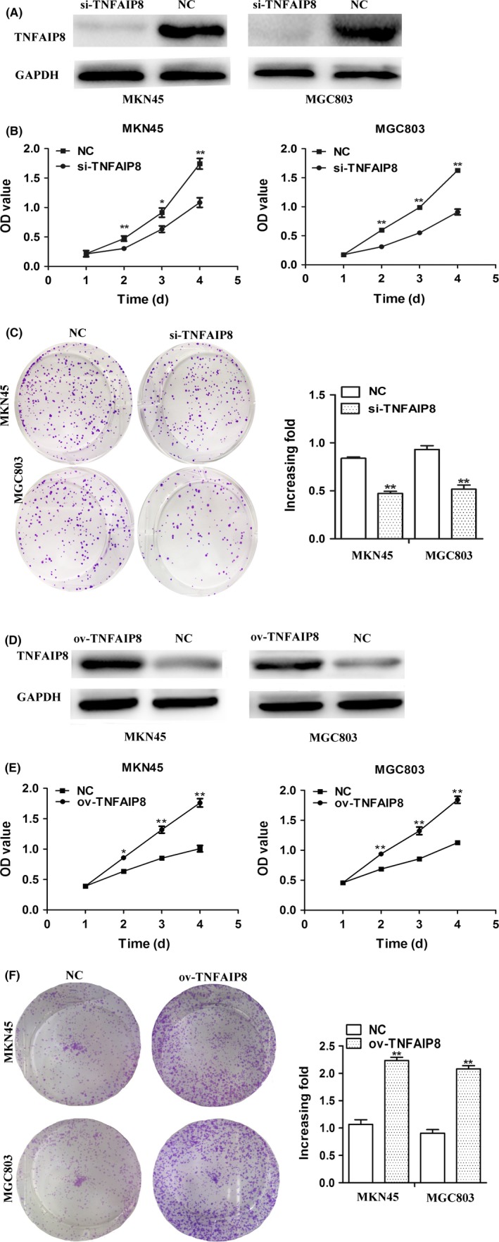
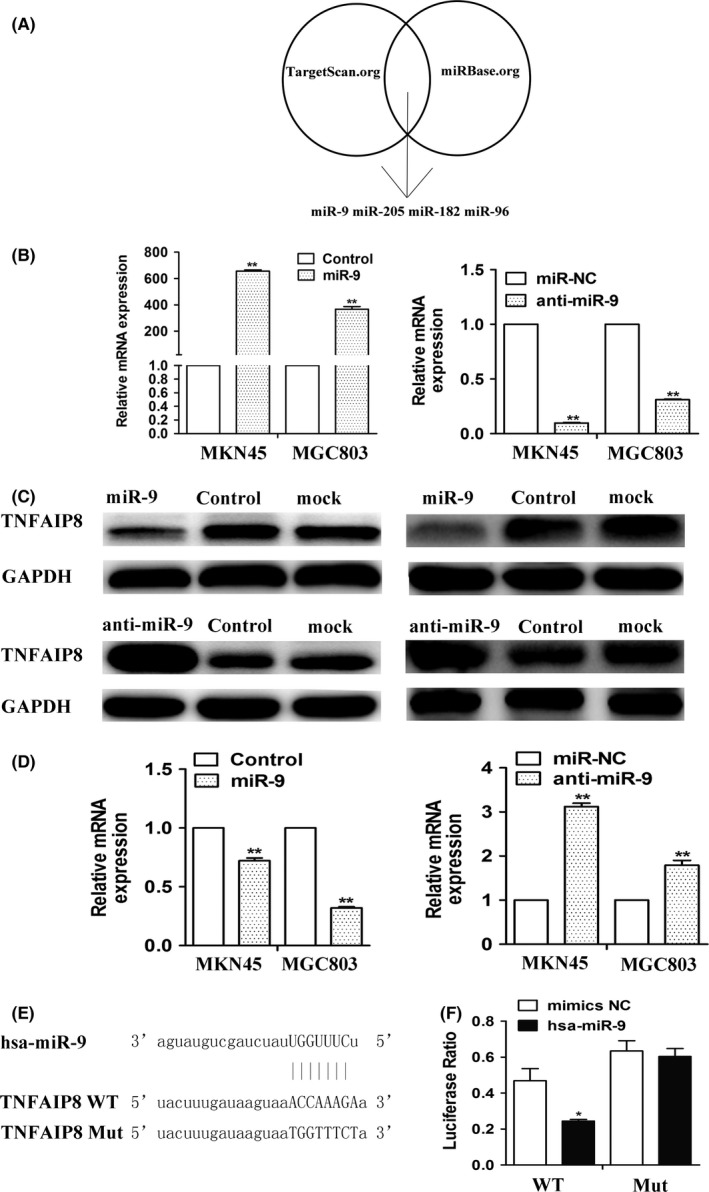
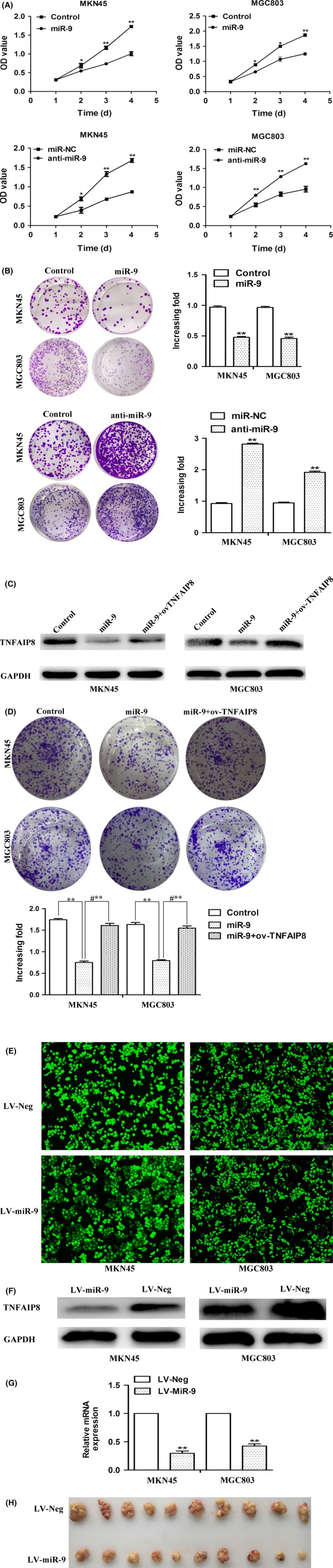
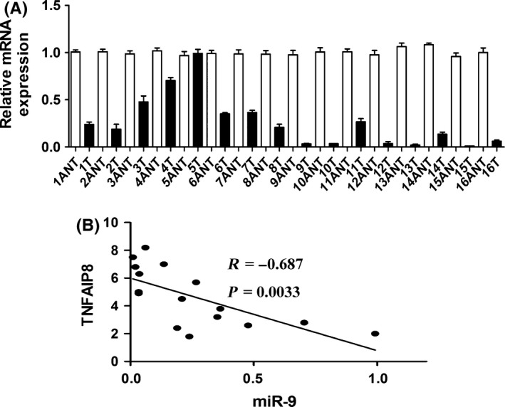
Materials and methods: Levels of miR-9 and TNFAIP8 expression in GC tissues and in human GC cell lines were studied using qualitative real-time PCR (qRT-PCR) and Western blotting. Cell viability was detected using the CCK-8 and clone formation assays. A dual-luciferase reporter system was used to confirm the target gene of miR-9.
Results: We found that the expression level of MicroRNA-9 in GC tissues and cell lines was significantly lower than that in adjacent non-cancerous tissues and human immortalized gastric epithelial cell (GES) line, respectively. In addition, overexpression of MicroRNA-9 markedly inhibited GC cell proliferation in vitro and tumour growth in vivo. Further experiments revealed that TNFAIP8 was a direct and functional target of MicroRNA-9 in GC and overexpression of MicroRNA-9 obviously down-regulated the expression of TNFAIP8, which was involved in the gastric carcinogenesis and cancer progression.
Conclusion: Our results suggested that MicroRNA-9-TNFAIP8 might represent a promising diagnostic biomarker for GC patients and could be a potential therapeutic target in the prevention and treatment of GC.
Keywords: TNFAIP8; biomarker; gastric cancer; miR-9; proliferation.
© 2017 John Wiley & Sons Ltd.
Conflict of interest statement
We have no conflicts of interest.
Figures
Similar articles
-
microRNA-665 is down-regulated in gastric cancer and inhibits proliferation, invasion, and EMT by targeting PPP2R2A.Cell Biochem Funct. 2020 Jun;38(4):409-418. doi: 10.1002/cbf.3485. Epub 2020 Jan 10. Cell Biochem Funct. 2020. PMID: 31923339
-
MicroRNA 495 Inhibits Proliferation and Metastasis and Promotes Apoptosis by Targeting Twist1 in Gastric Cancer Cells.Oncol Res. 2019 Feb 21;27(3):389-397. doi: 10.3727/096504018X15223159811838. Epub 2018 Apr 3. Oncol Res. 2019. Retraction in: Oncol Res. 2024 Sep 18;32(10):1681-1682. doi: 10.32604/or.2024.056898. PMID: 29615148 Free PMC article. Retracted.
-
MicroRNA-937 inhibits cell proliferation and metastasis in gastric cancer cells by downregulating FOXL2.Cancer Biomark. 2017 Dec 12;21(1):105-116. doi: 10.3233/CBM-170310. Cancer Biomark. 2017. Retraction in: Cancer Biomark. 2020;29(1):167. doi: 10.3233/CBM-200903. PMID: 29060929 Retracted.
-
Current research status of TNFAIP8 in tumours and other inflammatory conditions (Review).Int J Oncol. 2021 Jul;59(1):46. doi: 10.3892/ijo.2021.5226. Epub 2021 May 26. Int J Oncol. 2021. PMID: 34036374 Review.
-
Diverse functions of miR-328 in the carcinogenesis.Pathol Res Pract. 2023 Nov;251:154896. doi: 10.1016/j.prp.2023.154896. Epub 2023 Oct 16. Pathol Res Pract. 2023. PMID: 37852016 Review.
Cited by
-
TNFAIP8 promotes the proliferation and cisplatin chemoresistance of non-small cell lung cancer through MDM2/p53 pathway.Cell Commun Signal. 2018 Jul 31;16(1):43. doi: 10.1186/s12964-018-0254-x. Cell Commun Signal. 2018. PMID: 30064446 Free PMC article.
-
MicroRNA-205-5p inhibits skin cancer cell proliferation and increase drug sensitivity by targeting TNFAIP8.Sci Rep. 2021 Mar 11;11(1):5660. doi: 10.1038/s41598-021-85097-6. Sci Rep. 2021. Retraction in: Sci Rep. 2022 Sep 27;12(1):16103. doi: 10.1038/s41598-022-21189-1. PMID: 33707587 Free PMC article. Retracted.
-
TNFAIP8 promotes cell growth by regulating the Hippo pathway in epithelial ovarian cancer.Exp Ther Med. 2018 Dec;16(6):4975-4982. doi: 10.3892/etm.2018.6819. Epub 2018 Oct 2. Exp Ther Med. 2018. PMID: 30546405 Free PMC article.
-
MiR-9-5p Inhibits Glioblastoma Cells Proliferation Through Directly Targeting FOXP2 (Forkhead Box P2).Front Oncol. 2019 Nov 19;9:1176. doi: 10.3389/fonc.2019.01176. eCollection 2019. Front Oncol. 2019. PMID: 31824836 Free PMC article.
-
MiR-9 promotes tumorigenesis and angiogenesis and is activated by MYC and OCT4 in human glioma.J Exp Clin Cancer Res. 2019 Feb 22;38(1):99. doi: 10.1186/s13046-019-1078-2. J Exp Clin Cancer Res. 2019. PMID: 30795814 Free PMC article.
References
-
- Shen L, Shan YS, Hu HM, et al. Management of gastric cancer in Asia: resource‐stratified guidelines. Lancet Oncol. 2013;14:e535–e547. - PubMed
-
- Patel S, Wang FH, Whiteside TL, Kasid U. Identification of seven differentially displayed transcripts in human primary and matched metastatic head and neck squamous cell carcinoma cell lines: implications in metastasis and/or radiation response. Oral Oncol. 1997;33:197–203. - PubMed
-
- Yang M, Zhao Q, Wang X, et al. TNFAIP8 overexpression is associated with lymph node metastasis and poor prognosis in intestinal‐type gastric adenocarcinoma. Histopathology. 2014;65:517–526. - PubMed
MeSH terms
Substances
LinkOut - more resources
Full Text Sources
Other Literature Sources
Medical
Miscellaneous
