. 2017 Feb;26(2):227-241.
doi: 10.1002/pro.3072. Epub 2017 Jan 26.

The IDL of E. coli SSB links ssDNA and protein binding by mediating protein-protein interactions

Affiliations

The IDL of E. coli SSB links ssDNA and protein binding by mediating protein-protein interactions

Piero R Bianco et al. Protein Sci. 2017 Feb.

Abstract

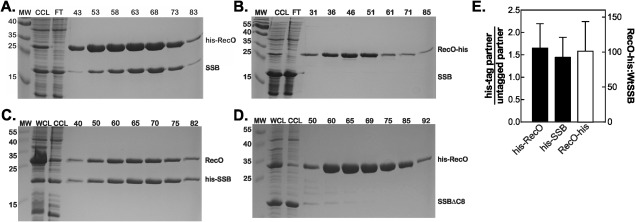
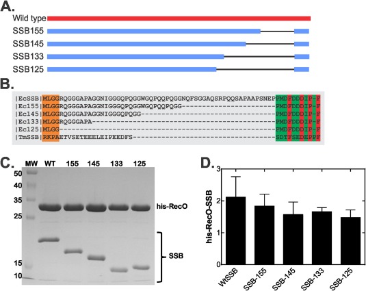
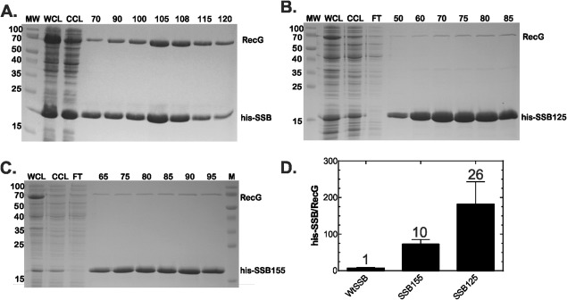
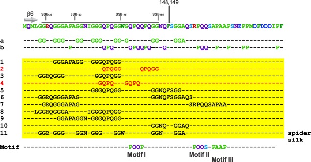
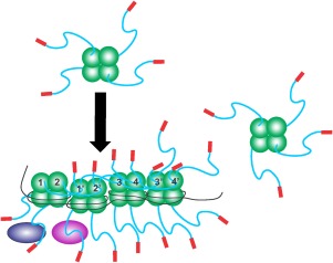
The E. coli single strand DNA binding protein (SSB) is essential to viability where it functions in two seemingly disparate roles: it binds to single stranded DNA (ssDNA) and to target proteins that comprise the SSB interactome. The link between these roles resides in a previously under-appreciated region of the protein known as the intrinsically disordered linker (IDL). We present a model wherein the IDL is responsible for mediating protein-protein interactions critical to each role. When interactions occur between SSB tetramers, cooperative binding to ssDNA results. When binding occurs between SSB and an interactome partner, storage or loading of that protein onto the DNA takes place. The properties of the IDL that facilitate these interactions include the presence of repeats, a putative polyproline type II helix and, PXXP motifs that may facilitate direct binding to the OB-fold in a manner similar to that observed for SH3 domain binding of PXXP ligands in eukaryotic systems.

Keywords: OB-fold; PXXP motif; RecG; RecO; SH3 domain; SSB.


Figures

Figure 1
Figure 1
SSB binds to RecO in vivo. The 1‐L cultures of cells expressing RecO and SSB were grown to early log phase, IPTG added to 500 µM and growth continued until early stationary phase. Cells were harvested by centrifugation, lysed and the cleared cell lysate applied to a 5‐mL nickel column as described in the Materials and Methods. Proteins were then eluted using an imidazole gradient following extensive washing to remove unbound proteins. Aliquots from various fractions throughout the purification were subjected to electrophoresis. The resulting Coomassie stained, 15% SDS‐PAGE gels of the purifications are presented. (A), hisRecO binds to SSB; (B), RecO‐his does not bind to SSB; (C), RecO binds to hisSSB; (D), formation of the RecO‐SSB complex requires the highly conserved and acidic SSB tip.; (E), Analysis of the gels in panels A–C. For each analysis, only the five central lanes of the peak were analyzed and the ratios averaged. MW, molecular weight marker; WCL, whole cell lysate; CCL, cleared cell lysate; FT, flow through; numbers, indicate fraction numbers from the peak elution profile. Proteins were eluted using a linear imidazole gradient from 30 to 500 mM.
Figure 2
Figure 2
SSB linker length affects binding to RecO. (A), Schematic of the wild type and linker domain mutants. The black line demarcates the region deleted in each mutant. (B), Sequence alignment of full length and linker domain mutants of E. coli SSB with T. maritima SSB. The alignment was done using PRALINE multiple sequence alignment and the result shows residues 112–178 (E. coli numbering)66. The green boxed region indicates the C‐terminal tail with invariant residues in red. (C) SSB linker domain mutants co‐elute with his‐RecO. The 100 mL cultures were grown to early log phase and expression induced with 500 µM IPTG. Cells were harvested in early stationary phase by centrifugation and the resulting pellets lysed. The cleared cell lysates were applied to 1mL nickel columns, extensively washed and eluted with elution buffer as described in the Materials and Methods. A Coomassie stained, 15% SDS‐PAGE gel with the apex fraction of each coelution is shown. To allow direct comparison, each lane was normalized to the amount of RecO loaded. (D), Quantification of SDS‐PAGE gels such as those shown in (C).
Figure 3
Figure 3
Alterations in the SSB linker eliminate binding to RecG. The 1‐L cultures of cells expressing RecG and SSB or linker mutants were grown to early log phase, IPTG added to 500 µM and growth continued until early stationary phase. Cells were harvested by centrifugation, lysed and the cleared cell lysate applied to a 5‐mL nickel column as described in the Materials and Methods. Proteins were then eluted using an imidazole gradient following extensive washing to remove unbound proteins. Aliquots from various fractions throughout the purification were subjected to electrophoresis. The resulting Coomassie stained, 12% SDS‐PAGE gels of the purifications are presented. (A) RecG binds to full‐length SSB as shown previously.7 (B) RecG does not bind appreciably to SSB125. (C) RecG does not bind appreciably to SSB155. MW, molecular weight marker; WCL, whole cell lysate; CCL, cleared cell lysate; FT, flow through; numbers, indicate fraction numbers from the peak elution profile.
Figure 4
Figure 4
The linker of EcSSB has a specialized sequence composition. The sequence of the C‐terminal 69 residues of EcSSB (top line) contains repetitive elements consisting of glycine, proline and glutamine. Sequence analysis of the protein was done using REPRO at http://www.ibi.vu.nl/programs/.38 The position of beta sheet 6 is indicated.34, 43 Lines a and b illuminate the over‐represented residues in the SSB C‐terminus (explained in the next). Lines 1–11 (yellow box) show a subset of the repeated sequence elements identified using REPRO.35, 38 The PXXP motifs were identified by eye. The amino acids spanning the putative PPII‐helix are indicated by the black line and the positions of the deletions in the linker mutants are also shown.
Figure 5
Figure 5
TmSSB forms stable complexes with TmRecG and EcRecO but not EcRecG. The 1‐L cultures of cells expressing RecG (Ec or Tm) with his‐TmSSB were grown to early log phase, IPTG added to 500 µM and growth continued until early stationary phase. Cells were harvested by centrifugation, lysed and the cleared cell lysate applied to a 5‐mL nickel column as described in the Materials and methods. Proteins were then eluted using an imidazole gradient following extensive washing to remove unbound proteins. Aliquots from various fractions throughout the purification were subjected to electrophoresis. Coomassie stained, 15% SDS‐PAGE gels showing various stages during the purification are presented. (A) TmRecG binds to TmSSB. (B) EcRecG does not bind appreciably to TmSSB. (C) TmSSB binds to EcRecO. (D), Analysis of the gels in panels A–C. The data for EcSSB/EcRecG are from column 1, Figure 3(D) and are presented here for comparison. MW, molecular weight marker; WCL, whole cell lysate; CCL, cleared cell lysate; numbers indicate fraction numbers from the peak elution profile.
Figure 6
Figure 6
The sequence composition of the EcSSB linker influences binding to RecO. (A) Schematic of EcSSB, five Mt‐Ec‐SSB hybrid proteins and MtSSB is shown. Residue numbering is from the E. coli sequence with methionine as 1. Numbers immediately above the hybrid proteins demarcates where the sequence changes from E. coli to M. tuberculosis. (B) Some hybrid proteins bind to EcRecO while others do not. Quantification of the Coomassie‐stained gels of apex fractions from each coelution of his‐RecO and the five hybrid SSB proteins is shown. The 100 mL cultures of his‐RecO and each of the five hybrid were grown separately to early log phase and expression induced with 500 µM IPTG. Cells were harvested in early stationary phase by centrifugation and the resulting pellets lysed. The cleared cell lysates were applied to 1‐mL nickel columns, extensively washed and eluted with elution buffer as described in the Materials and methods. Eluted fractions were subjected to electrophoresis in 15% SDS‐PAGE gels.
Figure 7
Figure 7
The proposed PPII helix region is important to EcSSB function. Homology modeling of the EM‐3 and 4 proteins was done at Swiss‐model in automated mode using PDB file 1QVC as a template.37, 39, 43 Models were built on the first subunit of 1QVC. As residues 1–111 in each model are identical to 1QVC they are not displayed. Instead, the Connolly surfaces of residues 110 to the C‐terminus of each model are shown with the N‐termini at the top left of each image.44, 67 For EcSSB this is to residue 145; for EM‐3 it extends to position 149, and for EM‐4 to amino acid 161. Colouring is done according to hydrophobicity as indicated and the orientation of each region is approximately the same. A sequence alignment of the hybrids to EcSSB is shown in Supporting Information Figure S2.
Figure 8
Figure 8
SSB linkers mediate all protein–protein interactions. Schematics of wild type SSB proteins binding to ssDNA and to partners. The colouring of SSB monomers is as follows the core domain in green, linker in blue and acidic tip in red. Here tetramers have their functional C‐terminal domains exposed in solution. Upon binding to ssDNA the IDLs of monomers 1 and 2, bind to the OB‐folds of monomers 1′ and 2′, respectively. Concurrently, the linkers of monomers 1′ and 2′ bind to the OB‐folds of monomers 3 and 4, and their IDLs bind to monomers 3′and 4′, respectively. The C‐termini of subunits 3′ and 4′ are available to bind to an incoming tetramer as shown on the right. On the opposite side of each tetramer, C‐termini are available for binding to interactome partners such as RecG or RecO (purple and blue ovals). For simplicity, SSB–SSB interactions are shown in the top subunits only, and SSB interactome partners binding in the lower subunits.

Similar articles

Cited by

References

    1. Shereda RD, Kozlov AG, Lohman TM, Cox MM, Keck JL (2008) SSB as an organizer/mobilizer of genome maintenance complexes. Crit Rev Biochem Mol Biol 43:289–318. - PMC - PubMed
    1. Chase JW, Williams KR (1986) Single‐stranded DNA binding proteins required for DNA replication. Annu Rev Biochem 55:103–136. - PubMed
    1. Meyer RR, Laine PS (1990) The single‐stranded DNA‐binding protein of Escherichia coli . Microbiol Rev 54:342–380. - PMC - PubMed
    1. Kowalczykowski SC, Dixon DA, Eggleston AK, Lauder SD, Rehrauer WM (1994) Biochemistry of homologous recombination in Escherichia coli . Microbiol Rev 58:401–465. - PMC - PubMed
    1. Lohman T, Ferrari M (1994) Escherichia coli single‐stranded DNA‐binding protein: multiple DNA‐binding modes and cooperativities. Ann Rev Biochem 63:527–570. - PubMed

MeSH terms