

Hitchhiker's Guide to Voltammetry: Acute and Chronic Electrodes for in Vivo Fast-Scan Cyclic Voltammetry
- PMID: 28127962
- PMCID: PMC5783156
- DOI: 10.1021/acschemneuro.6b00393
Hitchhiker's Guide to Voltammetry: Acute and Chronic Electrodes for in Vivo Fast-Scan Cyclic Voltammetry
Abstract
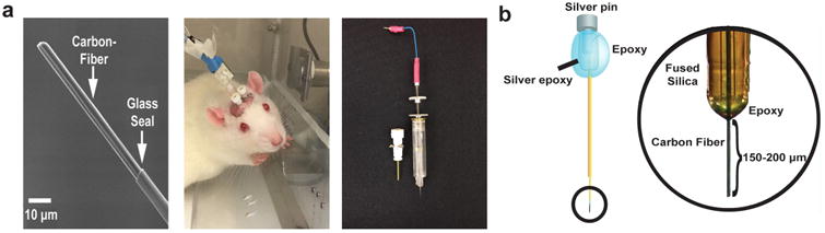
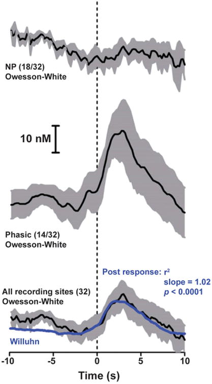
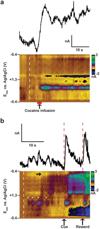
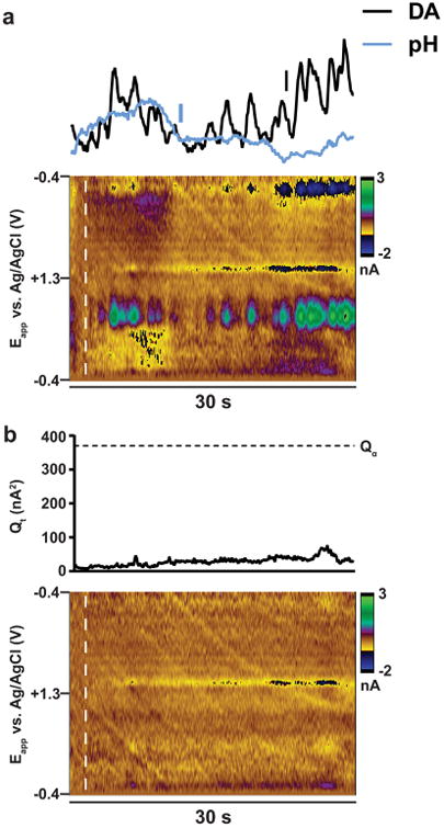
Fast-scan cyclic voltammetry (FSCV) has been used for over 20 years to study rapid neurotransmission in awake and behaving animals. These experiments were first carried out with carbon-fiber microelectrodes (CFMs) encased in borosilicate glass, which can be inserted into the brain through micromanipulators and guide cannulas. More recently, chronically implantable CFMs constructed with small diameter fused-silica have been introduced. These electrodes can be affixed in the brain with minimal tissue response, which permits longitudinal measurements of neurotransmission in single recording locations during behavior. Both electrode designs have been used to make novel discoveries in the fields of neurobiology, behavioral neuroscience, and psychopharmacology. The purpose of this Review is to address important considerations for the use of FSCV to study neurotransmitters in awake and behaving animals, with a focus on measurements of striatal dopamine. Common issues concerning experimental design, data collection, and calibration are addressed. When necessary, differences between the two methodologies (acute vs chronic recordings) are discussed. The topics raised in this Review are particularly important as the field moves beyond dopamine toward new neurochemicals and brain regions.
Keywords: Fast-scan cyclic voltammetry; carbon-fiber microelectrodes; chemometrics; dopamine; principal component regression.
Figures
Similar articles
-
Sampling phasic dopamine signaling with fast-scan cyclic voltammetry in awake, behaving rats.Curr Protoc Neurosci. 2015 Jan 5;70:7.25.1-7.25.20. doi: 10.1002/0471142301.ns0725s70. Curr Protoc Neurosci. 2015. PMID: 25559005 Free PMC article.
-
Improving in Situ Electrode Calibration with Principal Component Regression for Fast-Scan Cyclic Voltammetry.Anal Chem. 2018 Nov 20;90(22):13434-13442. doi: 10.1021/acs.analchem.8b03241. Epub 2018 Oct 29. Anal Chem. 2018. PMID: 30335966
-
A pipette-based calibration system for fast-scan cyclic voltammetry with fast response times.Biotechniques. 2016 Nov 1;61(5):269-271. doi: 10.2144/000114476. eCollection 2016. Biotechniques. 2016. PMID: 27839513
-
Electrochemical Analysis of Neurotransmitters.Annu Rev Anal Chem (Palo Alto Calif). 2015;8:239-61. doi: 10.1146/annurev-anchem-071114-040426. Epub 2015 May 4. Annu Rev Anal Chem (Palo Alto Calif). 2015. PMID: 25939038 Free PMC article. Review.
-
In Vivo Electrochemical Sensors for Neurochemicals: Recent Update.ACS Sens. 2019 Dec 27;4(12):3102-3118. doi: 10.1021/acssensors.9b01713. Epub 2019 Nov 25. ACS Sens. 2019. PMID: 31718157 Review.
Cited by
-
Sub-second Dopamine and Serotonin Signaling in Human Striatum during Perceptual Decision-Making.Neuron. 2020 Dec 9;108(5):999-1010.e6. doi: 10.1016/j.neuron.2020.09.015. Epub 2020 Oct 12. Neuron. 2020. PMID: 33049201 Free PMC article.
-
Perspective-Assessing Electrochemical, Aptamer-Based Sensors for Dynamic Monitoring of Cellular Signaling.ECS Sens Plus. 2023 Dec 1;2(4):042401. doi: 10.1149/2754-2726/ad15a1. Epub 2023 Dec 27. ECS Sens Plus. 2023. PMID: 38152504 Free PMC article.
-
Closed-Loop Implantable Therapeutic Neuromodulation Systems Based on Neurochemical Monitoring.Front Neurosci. 2019 Aug 20;13:808. doi: 10.3389/fnins.2019.00808. eCollection 2019. Front Neurosci. 2019. PMID: 31481864 Free PMC article. Review.
-
A ratiometric photoelectrochemical microsensor based on a small-molecule organic semiconductor for reliable in vivo analysis.Chem Sci. 2021 Sep 1;12(39):12977-12984. doi: 10.1039/d1sc03069h. eCollection 2021 Oct 13. Chem Sci. 2021. PMID: 34745528 Free PMC article.
-
Polymeric Composite-Based Electrochemical Sensing Devices Applied in the Analysis of Monoamine Neurotransmitters.Biosensors (Basel). 2025 Jul 9;15(7):440. doi: 10.3390/bios15070440. Biosensors (Basel). 2025. PMID: 40710090 Free PMC article. Review.
References
-
- Nieoullon A, Cheramy A, Glowinski J. Nigral and striatal dopamine release under sensory stimuli. Nature. 1977;269:340–342. - PubMed
-
- Cheramy A, Leviel V, Glowinski J. Dendritic release of dopamine in the substantia nigra. Nature. 1981;289:537–542. - PubMed
-
- Delgado JM, DeFeudis FV, Roth RH, Ryugo DK, Mitruka BM. Dialytrode for long term intracerebral perfusion in awake monkeys. Arch Int Pharmacodyn Ther. 1972;198:9–21. - PubMed
-
- Ungerstedt U, Pycock C. Functional correlates of dopamine neurotransmission. Bull Schweiz Akad Med Wiss. 1974;30:44–55. - PubMed
-
- Zetterstrom T, Sharp T, Marsden CA, Ungerstedt U. In vivo measurement of dopamine and its metabolites by intracerebral dialysis: changes after d-amphetamine. J Neurochem. 1983;41:1769–1773. - PubMed
Publication types
MeSH terms
Grants and funding
LinkOut - more resources
Full Text Sources
Other Literature Sources
