. 2018 Sep;38(9):1654-1663.
doi: 10.1177/0271678X17690164. Epub 2017 Jan 27.

Different types of white matter hyperintensities in CADASIL: Insights from 7-Tesla MRI

Affiliations

Different types of white matter hyperintensities in CADASIL: Insights from 7-Tesla MRI

François De Guio et al. J Cereb Blood Flow Metab. 2018 Sep.

Abstract

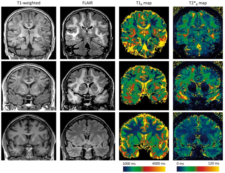
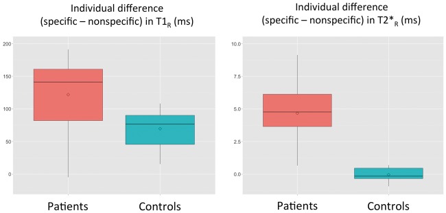
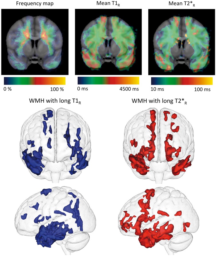
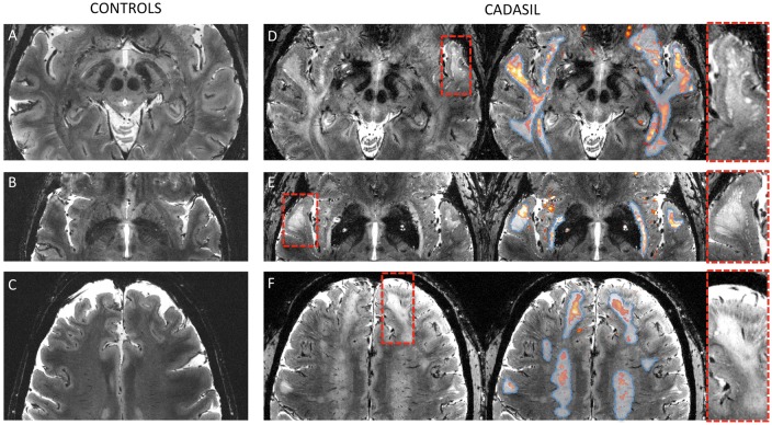
In Cerebral Autosomal Dominant Arteriopathy with Subcortical Infarcts and Leukoencephalopathy (CADASIL), by contrast to sporadic cerebral small vessel disease related to age and hypertension, white matter hyperintensities (WMH) are frequently observed in the white matter of anterior temporal poles, external capsules, and superior frontal regions. Whether these WMH (specific WMH) differ from those observed in other white matter areas (nonspecific WMH) remains unknown. Twenty patients were scanned to compare specific and nonspecific WMH using high-resolution images and analyses of relaxation times (T1R: longitudinal relaxation time and T2*R: effective transversal relaxation time). Specific WMH were characterized by significantly longer T1R and T2*R (T1R: 2309 ± 120 ms versus 2145 ± 138 ms; T2*R: 40 ± 5 ms versus 35 ± 5 ms, p < 0.001). These results were not explained by the presence of dilated perivascular spaces found in the close vicinity of specific WMH. They were not either explained by the normal regional variability of T1R and T2*R in the white matter nor by systematic imaging artifacts as shown by the study of 17 age- and sex-matched healthy controls. Our results suggest large differences in water content between specific and nonspecific WMH in CADASIL, supporting that mechanisms underlying WMH may differ according to their location.

Keywords: CADASIL; cerebral small vessel disease; dilated perivascular spaces; relaxometry; white matter hyperintensities.


Figures

Figure 1.
Figure 1.
Spatial heterogeneity of T1R and T2*R within white matter hyperintensities. Three tesla coronal slices of 3D T1-weighted, 3D FLAIR images and 7 T reconstructed T1R and T2*R maps of a 65-year CADASIL patient with extensive white matter hyperintensities (WMH) (first and second lines) and of a 60-year control (third line). (The 3D FLAIR has been acquired during an ongoing MRI protocol and is used here solely for illustration purpose.) T1-weighted and FLAIR images show WMH both in deep white matter and in specific regions such as temporal poles, external capsules, and superior frontal areas. Relaxometry maps illustrate that WMH with the longest relaxation times are located in anterior temporal, subinsular, and superior frontal areas. From deep white matter to these areas, we can observe an increase in T2*R within WMH from 30 ms to more than 120 ms. By comparison, T2*R is much shorter and homogeneous in the white matter of the control (around 25 ms). CADASIL: Cerebral Autosomal Dominant Arteriopathy with Subcortical Infarcts and Leukoencephalopathy; FLAIR: Fluid-attenuated inversion recovery; MRI: magnetic resonance imaging.
Figure 2.
Figure 2.
Boxplots illustrating the mean differences in T1R and T2*R between specific and nonspecific white matter hyperintensities calculated for each CADASIL patient and between corresponding ROI containing normal appearing white matter calculated for each control. Differences in T1R and T2*R were computed for each patient between voxels corresponding to specific and nonspecific white matter hyperintensities (WMH). Differences in T1R and T2*R were computed for each control between voxels of the specific ROI and voxels of the nonspecific ROI containing normal appearing white matter. The boxplots represent mean values and spread at the group level of these differences. Please note that T1R and T2*R are represented using different scales. CADASIL: Cerebral Autosomal Dominant Arteriopathy with Subcortical Infarcts and Leukoencephalopathy; ROI: region of interest.
Figure 3.
Figure 3.
Group analysis of spatial heterogeneity of T1R and T2*R within white matter hyperintensities. Top (from left to right): Frequency map (for each voxel, proportion of patients presenting with white matter hyperintensities (WMH) for this given voxel), T1R map and T2*R map in MNI template across the CADASIL group (N = 20) showing WMH with long relaxation times in juxta-cortical areas (anterior temporal poles and superior frontal gyri). Bottom: Segmentation of WMH characterized by long T1R (left) or long T2*R (right) values (two standard deviations above the mean values calculated on all voxels of WMH in CADASIL patients). WMH in these fronto-temporal regions rigorously match specific WMH. CADASIL: Cerebral Autosomal Dominant Arteriopathy with Subcortical Infarcts and Leukoencephalopathy.
Figure 4.
Figure 4.
Ultra-high resolution MRI of specific white matter hyperintensities (WMH). Native 7 T ultra-high resolution (0.25 × 0.25 × 1 mm3) structural T2*-weighted images of two controls (a: 59 years; b and c: 60 years) and two CADASIL patients (d: 57 years, e and f: 61 years). In patients (right column), T2*R maps (1 × 1 × 1 mm3) were overlaid (red to yellow color) after registration and segmentation (T2*R > 50 ms) and whole white matter hyperintensities (WMH) obtained from conventional FLAIR sequences were delineated (dashed blue line). Red dashed boxes were zoomed in to show the unique aspect of the border between the cortical mantle and underlying white matter. Specific WMH are characterized by long T2*R in the close vicinity of which multiple dilated perivascular spaces were observed at the cortico-subcortical junction. Otherwise, no macroscopic visible abnormality was found in areas directly corresponding to long T2*R. In controls, T2*R was much shorter and there were few if any dilated perivascular spaces detectable. CADASIL: Cerebral Autosomal Dominant Arteriopathy with Subcortical Infarcts and Leukoencephalopathy; FLAIR: Fluid-attenuated inversion recovery.

References

    1. Chabriat H, Joutel A, Dichgans M, et al. Lancet Neurol 2009; 8: 643–653. - PubMed
    1. Auer DP, Putz B, Gossl C, et al. Differential lesion patterns in CADASIL and sporadic subcortical arteriosclerotic encephalopathy: MR imaging study with statistical parametric group comparison. Radiology 2001; 218: 443–451. - PubMed
    1. O’Sullivan M, Jarosz JM, Martin RJ, et al. MRI hyperintensities of the temporal lobe and external capsule in patients with CADASIL. Neurology 2001; 56: 628–634. - PubMed
    1. Iadecola C. The pathobiology of vascular dementia. Neuron 2013; 80: 844–866. - PMC - PubMed
    1. Duering M, Csanadi E, Gesierich B, et al. Incident lacunes preferentially localize to the edge of white matter hyperintensities: insights into the pathophysiology of cerebral small vessel disease. Brain 2013; 136: 2717–2726. - PubMed

Publication types

LinkOut - more resources