Observational Study
. 2017 Jan 27:8:14155.
doi: 10.1038/ncomms14155.

Disruption of cardiac cholinergic neurons enhances susceptibility to ventricular arrhythmias

Affiliations
Observational Study

Disruption of cardiac cholinergic neurons enhances susceptibility to ventricular arrhythmias

Christiane Jungen et al. Nat Commun. .

Abstract

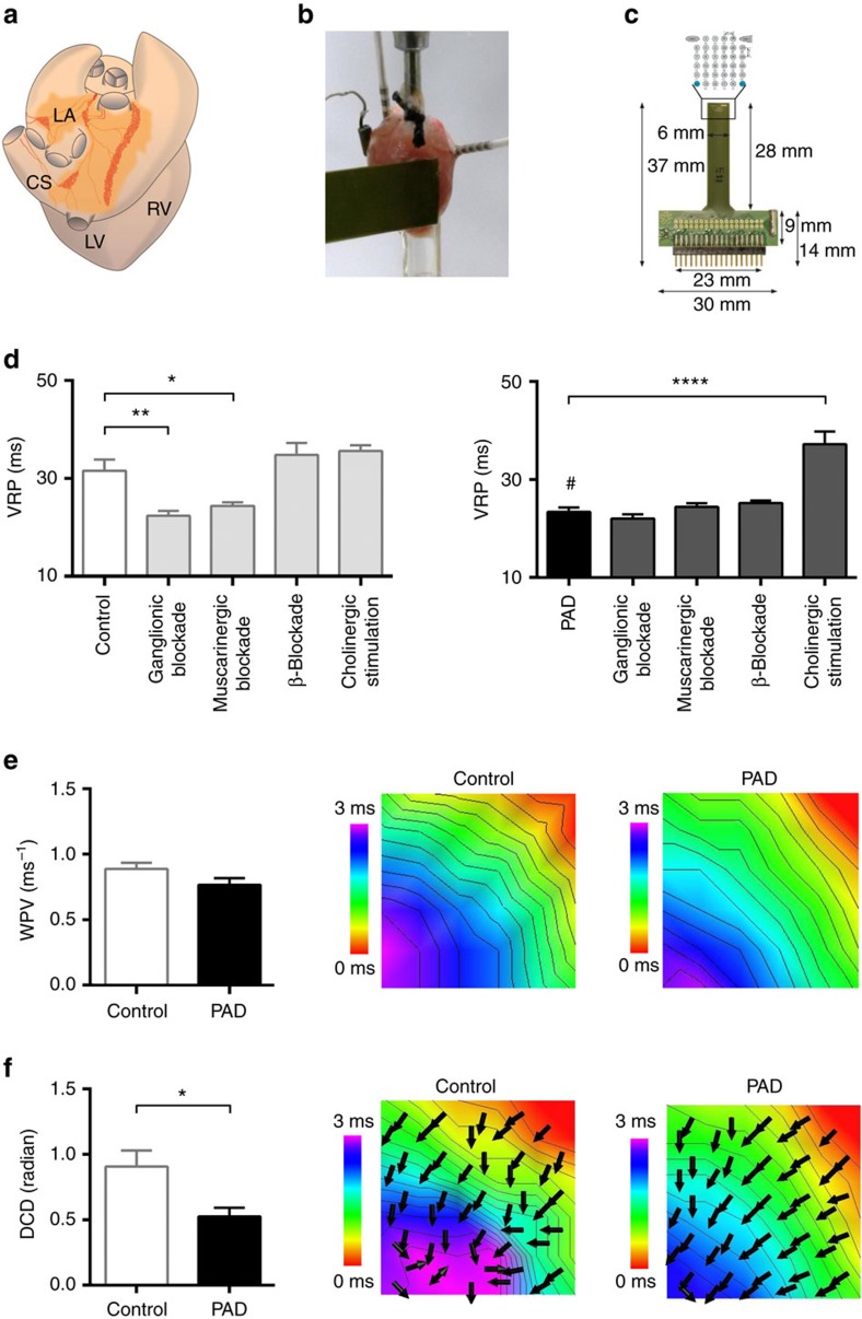
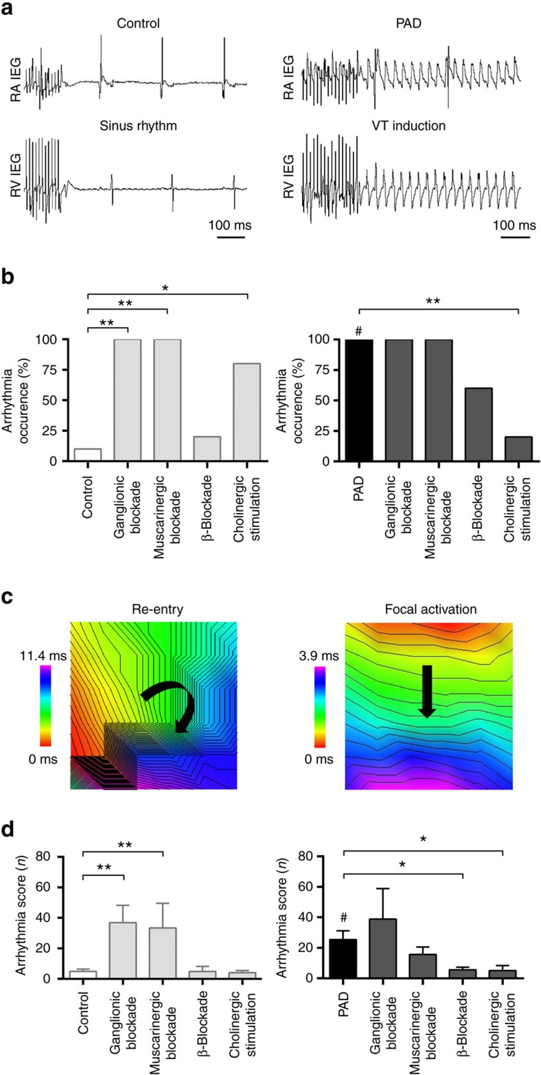
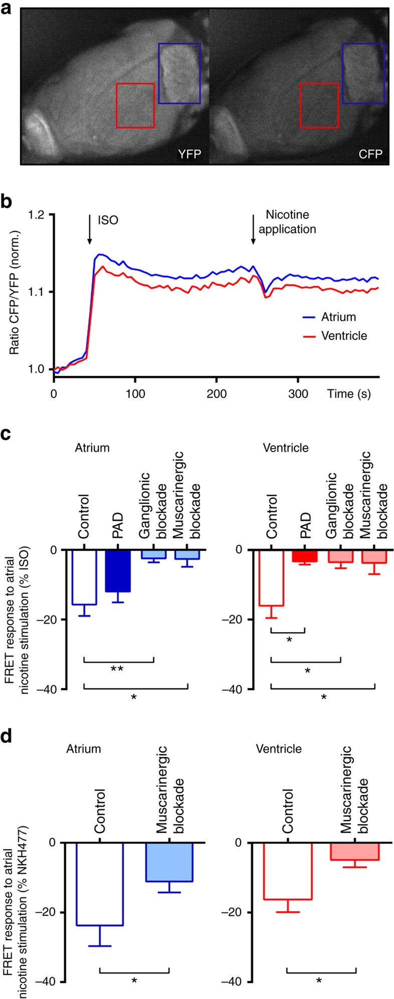
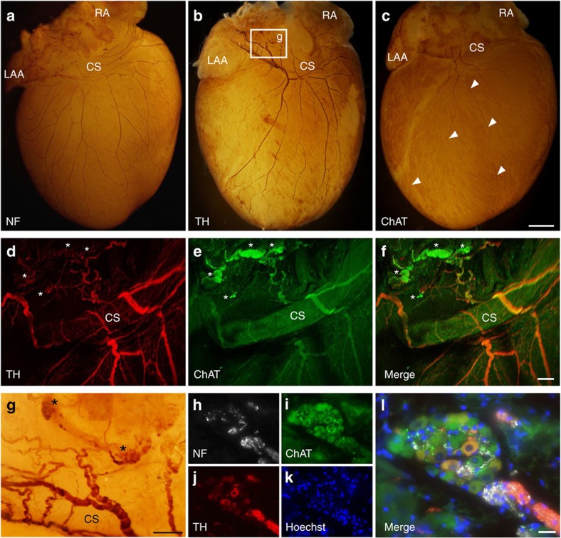
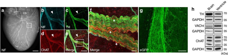
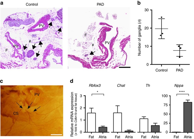
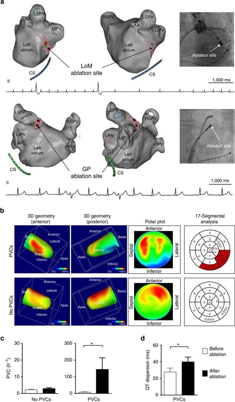
The parasympathetic nervous system plays an important role in the pathophysiology of atrial fibrillation. Catheter ablation, a minimally invasive procedure deactivating abnormal firing cardiac tissue, is increasingly becoming the therapy of choice for atrial fibrillation. This is inevitably associated with the obliteration of cardiac cholinergic neurons. However, the impact on ventricular electrophysiology is unclear. Here we show that cardiac cholinergic neurons modulate ventricular electrophysiology. Mechanical disruption or pharmacological blockade of parasympathetic innervation shortens ventricular refractory periods, increases the incidence of ventricular arrhythmia and decreases ventricular cAMP levels in murine hearts. Immunohistochemistry confirmed ventricular cholinergic innervation, revealing parasympathetic fibres running from the atria to the ventricles parallel to sympathetic fibres. In humans, catheter ablation of atrial fibrillation, which is accompanied by accidental parasympathetic and concomitant sympathetic denervation, raises the burden of premature ventricular complexes. In summary, our results demonstrate an influence of cardiac cholinergic neurons on the regulation of ventricular function and arrhythmogenesis.


Conflict of interest statement

The authors declare no competing financial interests.

Figures

Figure 1
Figure 1. Disruption of atrial cholinergic control modifies ventricular electrophysiology.
(a) Schematic drawing of a murine heart in posterior view. Light and dark orange surfaces depict areas of atrial fat with ganglionated plexi, which have been mechanically removed in partial atrial denervation (PAD) experiments. CS, coronary sinus; LA, left atrium; LV, left ventricle; RV, right ventricle. (b) Example of a murine heart within the Langendorff setup. Intracardial and epicardial catheters, as well as the epicardial multi-electrode array, are depicted. (c) The epicardial multi-electrode array is depicted with an enlargement of the schematic electrode layout. (d) The ventricular refractory periods (VRPs) are presented for control and PAD hearts with different pharmacological interventions. In comparison with control (n=10), the VRP was decreased by PAD (denoted by #; n=10; P=0.003; unpaired t-test), by ganglionic blockade (hexamethonium, 5 × 10⁻⁴ M; n=5; P=0.002; Mann–Whitney test) and by muscarinergic blockade (atropine, 1 × 10⁻⁵ M; n=5; P=0.045; unpaired t-test). β-Blockade (propranolol, 1 × 10⁻⁶ M; n=5) and cholinergic stimulation (acetylcholine, 1 × 10⁻⁵ M; n=5) did not influence VRPs in controls. After PAD ganglionic, muscarinergic or β-blockade did not reduce the VRP. However, cholinergic stimulation in PAD hearts raised the VRP (n=5; P<0.0001; unpaired t-test) suggesting reversibility of PAD-induced shortening of VRPs. (e) Wave propagation velocity (WPV) was not influenced by PAD as depicted in the colour-coded reconstruction with isochrones (2 m s−1 distance between isochrones) of epicardial multi-electrode activation mapping. (f) Dispersion of conduction direction (DCD) was smaller after PAD (n=16) compared with controls (n=19; P=0.026; Mann–Whitney test). Note the more homogeneous arrow alignment in the right image. All the values shown are mean±s.e.m. *P<0.05, **P<0.01, ****P<0.0001.
Figure 2
Figure 2. Disruption of atrial cholinergic control increases ventricular arrhythmogenesis.
(a) Example of arrhythmia susceptibility testing using burst stimulation in the right ventricle (RV; cycle length 10 ms, duration 5 s.) without (left panel: control) or with (right panel: partial atrial denervation (PAD)) induction of a ventricular tachycardia (VT). Arrhythmias occurred more frequently in PAD hearts. RA, right atrium; IEG, intracardiac electrogram. (b) Susceptibility to ventricular arrhythmias increased after PAD (right panel, black bar, 100%, n=10) compared with control hearts (left panel, white bar, 10%, n=10; denoted by #, P<0.0001). In control hearts, ganglionic (hexamethonium, 5 × 10⁻⁴ M; n=5; P=0.002) or muscarinergic blockade (atropine, 1 × 10⁻⁵ M, n=5; P=0.002) increased arrhythmia susceptibility. β-Blockade (propranolol, 1 × 10⁻⁶ M; n=5) did not significantly influence arrhythmia inducibility in controls. Cholinergic stimulation (acetylcholine, 1 × 10⁻⁵ M, n=5; P=0.017) led to an increase in arrhythmia susceptibility in control hearts in line with previous findings. In PAD hearts, ganglionic (n=5), muscarinergic (n=5) or β-blockade (n=5) did not significantly influence arrhythmia inducibility. Cholinergic stimulation reduced VT occurrence in PAD hearts (n=5; P=0.004). Fisher's exact test was used for all analyses in b. (c) Examples of ventricular arrhythmias with different underlying mechanisms (left: re-entry; right: focal). Red colour displays areas of earliest activation, purple of latest activation. (d) The arrhythmia score classifies the induced ventricular arrhythmias. After PAD, ventricular arrhythmias tended to be longer and more severe compared with controls (denoted by #; n=10; P=0.003; unpaired t-test), which is reflected by higher score values. In control hearts, ganglionic (n=5; P=0.002; unpaired t-test) or muscarinergic blockade (n=5; P=0.010; Mann–Whitney test) increased the arrhythmia score, while it was not affected by β-blockade or cholinergic stimulation. In PAD hearts, ganglionic (n=5) or muscarinergic blockade (n=5) did not alter the arrhythmia score. β-Blockade (n=5; P=0.011; Mann–Whitney test) or cholinergic stimulation (n=5; P=0.036; unpaired t-test) reduced it, indicating that PAD or pharmacological modulation of the cardiac autonomic tone impacts arrhythmia susceptibility. All the values shown are mean±s.e.m. *P<0.05, **P<0.01.
Figure 3
Figure 3. Atrial cholinergic modulation alters ventricular cAMP levels.
(a) Cyclic adenosine monophosphate (cAMP) biosensor fluorescence in yellow (YFP) and cyan (CFP) channels during FRET measurements in a Langendorff-perfused heart. The red/blue squares at the ventricle/atrium indicate the areas of measurement. (b) Representative experiment depicting time-resolved cAMP dynamics (presented as normalized CFP/YFP ratio) during perfusion with isoproterenol (ISO, 1 × 10⁻⁷ M) and targeted atrial application of nicotine (6.2 × 10⁻⁶ M). cAMP levels in the left atrium and ventricle were decreased by nicotine. (c) Relative changes in cAMP levels (% of ISO response) in atria and ventricles during local atrial application of nicotine in control hearts without pharmacological intervention (n=8), after partial atrial denervation (PAD; n=5) and during ganglionic (hexamethonium, 5 × 10⁻⁴ M; n=5) or muscarinergic blockade (atropine, 1 × 10⁻⁵ M; n=5) are depicted. In the atrium, targeted atrial nicotine application reduced relative atrial cAMP levels in controls and PAD hearts. This effect was reduced after ganglionic blockade (n=5; P=0.009; unpaired t-test) or muscarinergic blockade (n=5; P=0.011; Mann–Whitney test). In the ventricle, relative changes in cAMP levels during targeted atrial application of nicotine was obvious in controls (n=8) but abolished in PAD hearts (n=5; P=0.018; unpaired t-test) and during ganglionic (n=5; P=0.023; unpaired t-test) or muscarinergic blockade (n=5; P=0.036; unpaired t-test), indicating an inhibition of parasympathetic activity. (d) After stimulation with the water-soluble forskolin analogue NKH477 (1 × 10⁻⁵ M; n=5), relative changes in cAMP levels (% of NKH477 response) during local atrial application of nicotine in control hearts were recorded for the atrium and ventricle. Muscarinergic blockade applied in the same hearts reduced cAMP levels in the atrium (P=0.019; paired t-test) and ventricle (P=0.039; paired t-test) of NKH477 stimulated hearts. All the values shown are mean±s.e.m. *P<0.05, **P<0.01.
Figure 4
Figure 4. Interconnectivity of the atrial and ventricular intracardiac neural network.
(ac) Representative whole-mount stainings against neurofilament (NF; a), tyrosine hydroxylase (TH; b) and choline acetyltransferase (ChAT; c). Fibres traversed from the posterior left atrium along the pulmonary veins and the coronary sinus (CS) to the apex of the ventricle. Arrow heads mark the course of two ChAT-positive fibres. CS, coronary sinus; LAA, left atrial appendage; RA, right atrium. (df) Confocal imaging of TH (d) and ChAT (e) whole-mount co-staining revealed interwoven parasympathetic and sympathetic fibres originating from the parasympathetic ganglia (marked with asterisks). (g) Magnification from b depicting TH-positive fibres originating from atrial ganglia (marked with asterisks) with a dense network entwining the CS. (hl) Immunohistochemical staining of atrial ganglia illustrated the predominance of ChAT-positive cells. Scale bars, 1 mm (ac), 200 μm (dg), 25 μm (hl).
Figure 5
Figure 5. Sympathetic and parasympathetic ventricular innervation.
(a) Neurofilament (NF) staining depicting the rich innervation of the ventricle. The square marks the location of the fibres shown in bf. (bf) Confocal imaging of ventricular NF (b), tyrosine hydroxylase (TH; c) and choline acetyltransferase (ChAT; d). The white arrow heads display a TH- but not ChAT-positive fibre. The greater magnification (f) emphasizes the tight entanglement of fibres, with the majority being TH-positive and a smaller amount of parallel running ChAT-positive fibres. Arrows depict ChAT-positive fibres. (g) Native enhanced green fluorescent protein (eGFP) fluorescence of ChATBAC-eGFP mice demonstrated a delicate interconnectivity between parasympathetic ventricular fibres and cardiac myocytes. (h) Western blot analysis of ventricular tissue, using antibodies against TH, ChAT and VAChT revealed bands at the same molecular weight as in the brain (positive control), indicating the presence of a cholinergic system in the ventricles. Scale bars 1 mm (a), 100 μm (be), 10 μm (f), 25 μm (g).
Figure 6
Figure 6. Quantification of cardiac ganglia after PAD.
(a) Haematoxylin and eosin (H&E)-stained sections of control (left image) and partial atrial denervated (PAD) hearts (right image) are depicted. Cholinergic ganglia are marked with arrows. In PAD hearts, a great amount of epicardial fat and the ganglia located within have been removed. F, fat; PV, pulmonary vein; #, posterior atrial wall. Scale bar, 300 μm. (b) The total number of ganglia counted in the H&E-stained sections is reduced by 61% in PAD hearts compared with controls (n=3; P=0.100; Mann–Whitney test). (c) Whole-mount staining against choline acetyltransferase (ChAT) depicting disrupted cholinergic nerve fibres (arrows) after PAD. CS, coronary sinus. Scale bar, 500 μm. (d) Gene expression analysis of atrial myocardial tissue and epicardial fat via qRT–PCR (n=7) revealed significant differences in neuronal mRNA levels between the epicardial fat and cardiac atria. Compared with atrial tissue Rbfox3 mRNA (P=0.011; paired t-test), Chat mRNA (P=0.063; Wilcoxon signed-rank test) and Th mRNA (P=0.078; Wilcoxon signed-rank test) seemed to be slightly more prominent in the epicardial fat. Atrial Nppa expression (representing atrial natriuretic peptide) confirms correct fat preparation as it is only expressed within the atria (n=7; P<0.0001; paired t-test). All the values shown are mean±s.e.m. *P<0.05, ****P<0.0001.
Figure 7
Figure 7. Atrial neuroablation affects ventricular electrophysiology.
(a) Examples of three-dimensional (3D) guided ablation at the endocardial ligament of Marshall region (LoM; upper panel) and left superior ganglionated plexi (GP) region (lower panel) induces premature ventricular complexes (PVCs) during catheter ablation of AF. Red dots (VisiTag points) illustrate the sites of radiofrequency ablation. Left lateral (left) and anterolateral (right) views are shown. Note the PVCs occurring during ablation depicted in the ECG. A circular mapping catheter is located within the left superior pulmonary vein (LSPV; lower panel). The fluoroscopy image at the right shows the position of the ablation catheter and the circular mapping catheter positioned within the left inferior pulmonary vein (LIPV; upper panel) demonstrating the close proximity between this vein, the coronary sinus and the ablation site. CS, coronary sinus; LA, left atrium; LAA, left atrial appendage; right inferior pulmonary vein, RIPV; right superior pulmonary vein, RSPV. Scale bar, each 1,000 ms. (b) I-123-MIBG SPECT imaging revealed a reduced sympathetic innervation at the inferior and inferolateral parts of the left ventricle after catheter ablation of AF in patients with an increased PVC burden or symptomatic palpitations after AF ablation (n=4). In those without an increase in PVCs after AF ablation (n=4), regional left ventricular sympathetic innervation was less impaired. The right panel displays mean summed defect score values, whereas red parts depict areas of reduced uptake (mean summed defect score for one segment ≥0.5). (c) The majority of patients had no increase of PVCs per hour (No PVCs, n=105). A subset of patients (PVCs, n=6; P=0.031; Wilcoxon signed-rank test) reported palpitations or shortness of breath after catheter ablation of AF concomitant with an increase in PVCs per hour, irrespective of AF recurrence. (d) QT dispersion after catheter ablation was increased in patients with an elevated PVC burden (n=6; P=0.011; paired t-test). All the values shown are mean±s.e.m. *P<0.05.

References

    1. Mills T. W. Rhythm and innervation of the heart of the sea-turtle. J. Anat. Physiol. 21, 1–20 (1886). - PMC - PubMed
    1. Wickramasinghe S. R. & Patel V. V. Local innervation and atrial fibrillation. Circulation 128, 1566–1575 (2013). - PMC - PubMed
    1. Savage N. Physiology: beating stroke. Nature 493, S12–S13 (2013). - PubMed
    1. Pappone C. et al.. Pulmonary vein denervation enhances long-term benefit after circumferential ablation for paroxysmal atrial fibrillation. Circulation 109, 327–334 (2004). - PubMed
    1. Katritsis D. G. et al.. Autonomic denervation added to pulmonary vein isolation for paroxysmal atrial fibrillation: a randomized clinical trial. J. Am. Coll. Cardiol. 62, 2318–2325 (2013). - PubMed

Publication types

MeSH terms