. 2017 Jan 27:8:14246.
doi: 10.1038/ncomms14246.

A non-canonical mismatch repair pathway in prokaryotes

Affiliations

A non-canonical mismatch repair pathway in prokaryotes

A Castañeda-García et al. Nat Commun. .

Abstract

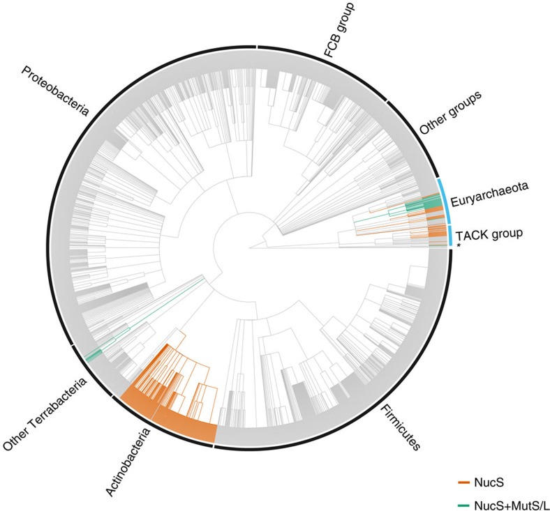
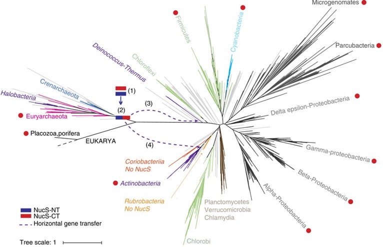
Mismatch repair (MMR) is a near ubiquitous pathway, essential for the maintenance of genome stability. Members of the MutS and MutL protein families perform key steps in mismatch correction. Despite the major importance of this repair pathway, MutS-MutL are absent in almost all Actinobacteria and many Archaea. However, these organisms exhibit rates and spectra of spontaneous mutations similar to MMR-bearing species, suggesting the existence of an alternative to the canonical MutS-MutL-based MMR. Here we report that Mycobacterium smegmatis NucS/EndoMS, a putative endonuclease with no structural homology to known MMR factors, is required for mutation avoidance and anti-recombination, hallmarks of the canonical MMR. Furthermore, phenotypic analysis of naturally occurring polymorphic NucS in a M. smegmatis surrogate model, suggests the existence of M. tuberculosis mutator strains. The phylogenetic analysis of NucS indicates a complex evolutionary process leading to a disperse distribution pattern in prokaryotes. Together, these findings indicate that distinct pathways for MMR have evolved at least twice in nature.


Conflict of interest statement

The authors declare no competing financial interests.

Figures

Figure 1
Figure 1. Identification and characterization of M. smegmatis NucS.
(a) Schematic representation of the process for identifying the nucS transposon mutant. ∼11,000 clones from the M. smegmatis insertion mutant library were replicated onto plates with (Rif) or without (No Rif) rifampicin (step 1). One single clone (circled) produced a high number of Rif-R colonies. After isolation and purification (step 2), the frequency of spontaneous Rif-R mutants (bottom plate) was checked and compared with that of the wild-type (upper plate), demonstrating its hypermutable phenotype. (b) Multiple sequence alignment of NucS sequences. M. tuberculosis (NucS_Mtu), M. smegmatis (NucS_Msm) and P. abyssi (NucS_Pab) sequences are from Uniprot (identifiers are P9WIY4, A0R1Z0 and Q9V2E8, respectively). Solid lines over the alignment indicate protein domains as defined previously for P. abyssi NucS (black, DNA-binding; grey, nuclease). Identical amino acid residues are shown in black. Catalytic residues required for nuclease activity in P. abyssi NucS are labelled with asterisks. Nuclease motifs of P. abyssi NucS are also indicated. (c) DNA-binding activity of NucS. In a gel-based EMSA, purified NucS protein (1–16 μM) is capable of binding to 45-mer ssDNA (50 nM) (left side) but not to 45-bp dsDNA (50 nM) (right side). The arrow indicates the position of the DNA–NucS complex.
Figure 2
Figure 2. Mutational effects of nucS deletion.
(a) Rates of spontaneous mutations conferring rifampicin, Rif-R (red), and streptomycin resistance, Str-R (grey) of M. smegmatis mc2 155 (WT), its ΔnucS derivative and the ΔnucS strain complemented with nucS from M. smegmatis mc2 155 (nucSSm). (b) Mutational spectrum of M. smegmatis mc2 155 (green) and its ΔnucS derivative (red). Bars represent the frequency of the types of change found in rpoB. (c) Rates of spontaneous mutations conferring Rif-R (red) and Str-R (grey) of S. coelicolor A3(2) M145 (WT), its ΔnucS derivative and the ΔnucS strain complemented with the wild-type nucS from S. coelicolor (nucSSco). Error bars represent 95% confidence intervals (n=20). Asterisks denote statistical significance (Likelihood ratio test under Luria-Delbruck model, Bonferroni corrected, P value <10−4 in all cases). Mutation rate: mutations per cell per generation.
Figure 3
Figure 3. Effect of nucS deletion on recombination.
(a) Chromosomal construct used to measure recombination between homologous or homeologous DNA sequences. The hyg gene is reconstituted by a single recombination event between two 517-bp overlapping fragments (striped), sharing different degree of sequence identity (100%, 95%, 90% and 85%) and separated by a kanamycin resistant (Kan-R) gene. Recombinant clones express hygromycin resistance and kanamycin susceptibility. (b) Rates of recombination between homologous and homeologous DNA sequences with different degree of identity (%) in M. smegmatis mc2 155 (WT, green squares) and its ΔnucS derivative (red diamonds). Error bars represent 95% confidence intervals (n=16). Asterisks denote statistical significance (Likelihood ratio test under Luria–Delbruck model, Bonferroni corrected, P value <10−4 in all cases).
Figure 4
Figure 4. Effects of NucS polymorphisms on mutation rates in the M. smegmatis ΔnucS surrogate model.
Rates of spontaneous mutations conferring Rif-R of the M. smegmatis ΔnucS complemented with wild-type nucSTBnucS/nucSTB; red) or containing each of the nine polymorphisms indicated (blue). Relative increases in mutation rates with respect to the control strain (ΔnucS/nucSTB; set to 1) are shown inside the column. Error bars represent 95% confidence intervals (n=20). Asterisks denote statistical significance (Likelihood ratio test under Luria-Delbruck model, Bonferroni corrected; ***P<0.001; **P<0.005). Mutation rate: mutations per cell per generation.
Figure 5
Figure 5. Phylogenetic profiling of NucS.
The NCBI taxonomic tree from 2,186 species from Bacteria (black outer label) and Archaea (blue outer label). Orange branches: NucS only; green branches: NucS and MutS–MutL. Bacteria includes Actinobacteria, Firmicutes, Proteobacteria, FCB (Fibrobacteres, Chlorobi and Bacteroidetes), Other Terrabacteria (Armatimonadetes, Chloroflexi, Cyanobacteria, Deinococcus-Thermus, Tenericutes and unclassified Terrabacteria) and other groups (Acidobacteria, Aquificae, Caldiserica, Chrysiogenetes, Deferribacteres, Dictyoglomi, Elusimicrobia, Fusobacteria, Nitrospirae, PVC group, Spirochaetes, Synergistetes, Thermodesulfobacteria, Thermotogae and unclassified bacteria). Archaea includes Euryarchaeota, TACK (Thaumarchaeota, Aigarchaeota, Crenarchaeota and Korarchaeota) and unclassified archaeal species (*). As NucS is absent in eukaryotes and viruses, these lineages were removed for clarity purposes. The tree was annotated using ggtree (http://www.bioconductor.org/packages/ggtree).
Figure 6
Figure 6. A model for NucS protein emergence and evolution.
The unrooted Tree of Life (available and based on ref. 50) was used to depict the proposed evolutionary history of NucS according to our data. The groups relevant to our model are highlighted. Coloured squares depict the NucS-NT (blue) and NucS-CT (red) terminal regions. This model proposes that NucS has an archaeal origin and emerged as a combination of two independent protein domains with complex evolutionary history. Numbers indicate the steps of the model: Both N-terminal and C-terminal regions likely emerged in the archaeal lineage (1). The CT region was transferred via HGT to very few Eukaryotes and to some Bacteria (main groups with any species having the NucS-CT region are labelled with red circles), where the CT domain combined with other regions outside the context of NucS. In the archaeal lineage, NT and CT regions fused to produce the full NucS (2). NucS expanded in many archaeal groups but was also lost in some others. The full NucS protein was transferred to Bacteria by at least two independent HGT events, one to some Deinoccocus-Thermus species (3) and another to Actinobacteria (4).

Similar articles

Cited by

References

    1. Friedberg E. et al. in DNA Repair and Mutagenesis 2nd edn American Society of Microbiology (2006).
    1. Eisen J. A. & Hanawalt P. C. A phylogenomic study of DNA repair genes, proteins, and processes. Mutat. Res. 435, 171–213 (1999). - PMC - PubMed
    1. Iyer R. R., Pluciennik A., Burdett V. & Modrich P. L. DNA mismatch repair: functions and mechanisms. Chem. Rev. 106, 302–323 (2006). - PubMed
    1. Matic I., Rayssiguier C. & Radman M. Interspecies gene exchange in bacteria: the role of SOS and mismatch repair systems in evolution of species. Cell 80, 507–515 (1995). - PubMed
    1. Sachadyn P. Conservation and diversity of MutS proteins. Mutat. Res. 694, 20–30 (2010). - PubMed

Publication types

MeSH terms