. 2017 Jan 27:7:36402.
doi: 10.1038/srep36402.

Pyrintegrin Induces Soft Tissue Formation by Transplanted or Endogenous Cells

Affiliations

Pyrintegrin Induces Soft Tissue Formation by Transplanted or Endogenous Cells

Bhranti S Shah et al. Sci Rep. .

Abstract

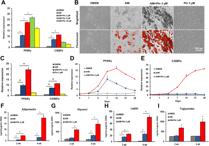
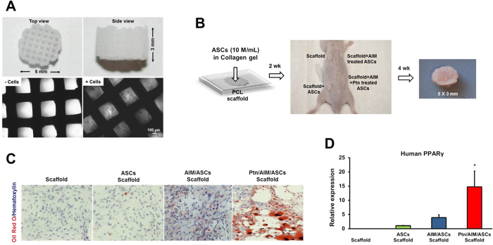
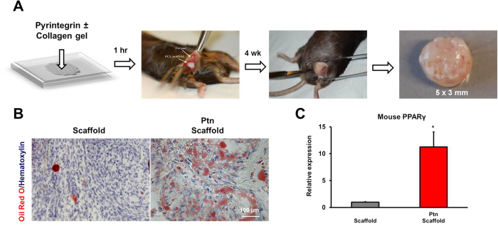
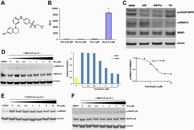
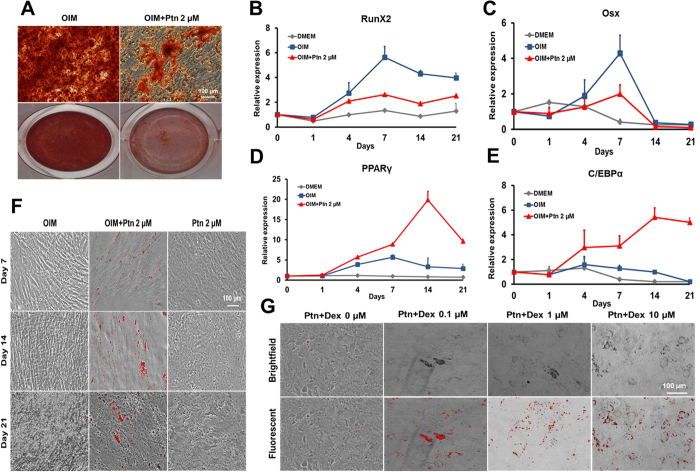
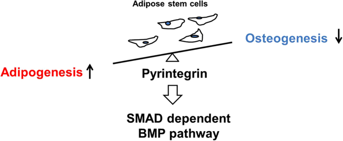
Focal adipose deficiency, such as lipoatrophy, lumpectomy or facial trauma, is a formidable challenge in reconstructive medicine, and yet scarcely investigated in experimental studies. Here, we report that Pyrintegrin (Ptn), a 2,4-disubstituted pyrimidine known to promote embryonic stem cells survival, is robustly adipogenic and induces postnatal adipose tissue formation in vivo of transplanted adipose stem/progenitor cells (ASCs) and recruited endogenous cells. In vitro, Ptn stimulated human adipose tissue derived ASCs to differentiate into lipid-laden adipocytes by upregulating peroxisome proliferator-activated receptor (PPARγ) and CCAAT/enhancer-binding protein-α (C/EBPα), with differentiated cells increasingly secreting adiponectin, leptin, glycerol and total triglycerides. Ptn-primed human ASCs seeded in 3D-bioprinted biomaterial scaffolds yielded newly formed adipose tissue that expressed human PPARγ, when transplanted into the dorsum of athymic mice. Remarkably, Ptn-adsorbed 3D scaffolds implanted in the inguinal fat pad had enhanced adipose tissue formation, suggesting Ptn's ability to induce in situ adipogenesis of endogenous cells. Ptn promoted adipogenesis by upregulating PPARγ and C/EBPα not only in adipogenesis induction medium, but also in chemically defined medium specifically for osteogenesis, and concurrently attenuated Runx2 and Osx via BMP-mediated SMAD1/5 phosphorylation. These findings suggest Ptn's novel role as an adipogenesis inducer with a therapeutic potential in soft tissue reconstruction and augmentation.


Conflict of interest statement

The authors declare no competing financial interests.

Figures

Figure 1
Figure 1. Pyrintegrin enhances adipogenesis of human adipose stem/progenitor cells in vitro.
(A) Ptn dose optimization by measuring PPARγ and C/EBPα expression in DMEM, AIM, AIM containing varying doses of Ptn 2-, 5- and 10-μM in hASC cultures after 4 days. Human GAPDH was used as the housekeeping gene and values representing mRNA of the DMEM group were defined as 1. AIM: adipogenesis induction medium; Ptn: Pyrintegrin. (B) Lipid accumulation in DMEM, AIM, AIM+Ptn 2-μM and Ptn 2-μM alone hASC cultures over 4 wks. Scale bar: 100 μm. (C) Relative mRNA expression of PPARγ and C/EBPα in DMEM, AIM, AIM+Ptn 2-μM and Ptn 2-μM alone hASC cultures at day 7. (D,E) Time-course real time RT-PCR analysis of PPARγ and C/EBPα expression of DMEM, AIM and AIM+Ptn 2-μM treated hASC cultures over 28 days. (F–I) Supernatant analysis of adiponectin, glycerol, leptin and triglycerides measured from DMEM, AIM and AIM+Ptn 2-μM treated hASC cultures at 2 and 4 wks. Data are expressed as mean ± S.D. *P < 0.05.
Figure 2
Figure 2. Pyrintegrin induces adipogenesis of transplanted human adipose stem/progenitor cells in vivo.
(A) Polycaprolactone (PCL) particles were melted and 3D-printed as cylinders, with top and side views and microscopic images without and with cells seeded in microchannels. (B) Infusion of human adipose derived stem cells (hASCs) in collagen gel into PCL microchannels, followed by implantation in the dorsum of athymic mice and retrieved in 4 wks, with a representative sample shown. (C) Representative histology images of in vivo retrieved samples stained for lipids by Oil-Red-O dye and nucleus by hematoxylin stain. (D) q RT-PCR analysis of human PPARγ, of in vivo retrieved samples. Scale bar: 100 μm. Data are expressed as mean ± S.D. *P < 0.05.
Figure 3
Figure 3. Pyrintegrin enhances endogenous adipose tissue formation in vivo.
(A) Pyrintegrin (Ptn) (10 μg/mL) was adsorbed in collagen gel that was infused in 3D-printed polycaprolactone (PCL) scaffold, followed by 1-h incubation at 37 °C and implantation in the inguinal fat pad of C57BL/6 mice. Representative sample image following scaffold retrieval after 4 wks. (B) Representative Oil-Red-O images of in vivo retrieved scaffold and Ptn-infused scaffold samples. (C) qRT-PCR analysis showing increased mouse PPARγ gene expression in Ptn-infused scaffold group. Scale bar: 100 μm. Data are expressed as means ± SD. *P < 0.05.
Figure 4
Figure 4. Pyrintegrin attenuates BMP-mediated SMAD activation.
(A) Ptn chemical structure. (B) Dose response of human PPPARγ luciferase assay. RLU, relative light units. (C) MAPK p44/p42 and SMAD1/5 phosphorylation by immunoblot after pretreatment with DMEM, AIM, AIM + Ptn and Ptn alone for 1 h. Equivalent protein loading confirmed by total SMAD1 and β-actin. (D) Western blot and densitometry analysis of SMAD1/5 phosphorylation following Ptn pretreatment for 30 min followed by treatment with BMP4 for 30 min. Equivalent protein loading confirmed by detection of total SMAD1 and β-actin. Hill plot of the inhibition of BMP4-stimulated SMAD1/5/8 phosphorylation by incubating hASCs with Pyrintegrin. (E) SMAD2 phosphorylation treated with BMP4 showing virtually negative Ptn effect on BMP4-mediated SMAD2 activity. (F) Ptn attenuated BMP4-mediated MAPK p38 phosphorylation modestly at concentrations ≥ 5 μM. Data are expressed as means ± SD. *P < 0.05.
Figure 5
Figure 5. Pyrintegrin attenuates osteogenic differentiation in vitro.
(A) Alizarin Red staining following treatment of adipose stem/progenitor cells (ASCs) in osteogenesis induction medium (OIM) and OIM+Pyrintegrin (Ptn) for 3 wks. (B,C) Time-course qPCR analysis of Runx2, Osx, PPARγ and C/EBPα mRNA expression measured over 21 days. (F) Lipid staining of hASCs treated with OIM, OIM+Ptn and Ptn alone at Days 7, 14 and 21. (G) Lipid staining of hASCs exposed to 2-μM Ptn and multiple dexamethasone doses (0, 0.1, 1 and 10 μM) for 21 days.
Figure 6
Figure 6. Proposed model for the role of Ptn signaling in hASCs.
See discussion for details.

Similar articles

Cited by

References

    1. Mao J. J. et al.. Facial reconstruction by biosurgery: cell transplantation versus cell homing. Tissue Eng Part B Rev 16, 257–262 (2010). - PMC - PubMed
    1. Stosich M. S. & Mao J. J. Adipose tissue engineering from human adult stem cells: clinical implications in plastic and reconstructive surgery. Plast Reconstr Surg 119, 71–83; discussion 84-75 (2007). - PMC - PubMed
    1. Hammer-Hansen N., Akram J. & Damsgaard T. E. The versatility of autologous fat transplantation in correction of facial deformities: a single-center experience. Plast Surg Int 2015, 703535 (2015). - PMC - PubMed
    1. Patrick C. W. Jr. Adipose tissue engineering: the future of breast and soft tissue reconstruction following tumor resection. Semin Surg Oncol 19, 302–311 (2000). - PubMed
    1. Patrick C. W. Jr. Tissue engineering strategies for adipose tissue repair. Anat Rec 263, 361–366 (2001). - PubMed

Publication types

MeSH terms