. 2017 Jan 27:7:41491.
doi: 10.1038/srep41491.

Protective effect of butin against ischemia/reperfusion-induced myocardial injury in diabetic mice: involvement of the AMPK/GSK-3β/Nrf2 signaling pathway

Affiliations

Protective effect of butin against ischemia/reperfusion-induced myocardial injury in diabetic mice: involvement of the AMPK/GSK-3β/Nrf2 signaling pathway

Jialin Duan et al. Sci Rep. .

Abstract

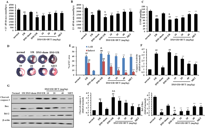
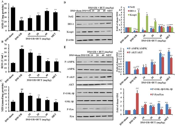
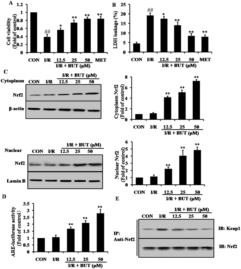
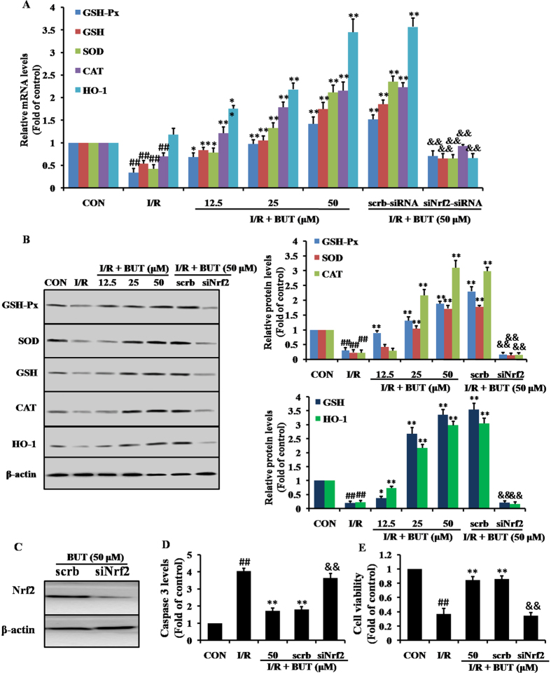
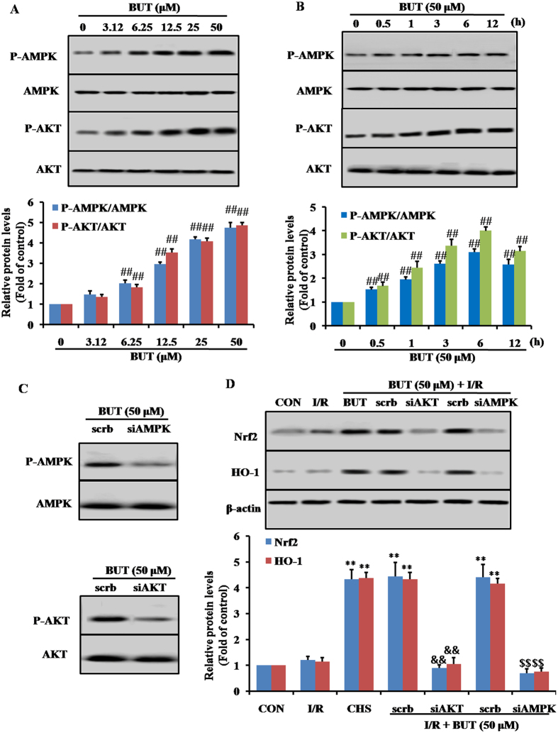
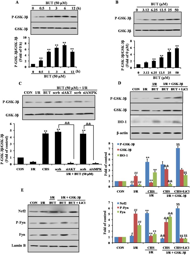
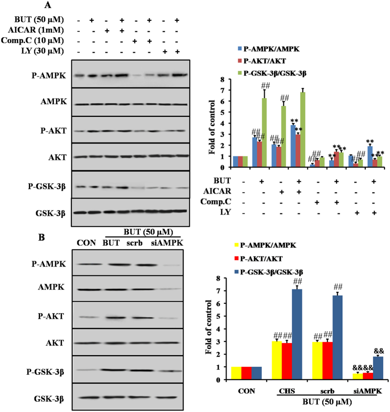
Hyperglycemia-induced reactive oxygen species (ROS) generation contributes to development of diabetic cardiomyopathy (DCM). This study was designed to determine the effect of an antioxidant butin (BUT) on ischemia/reperfusion-induced myocardial injury in diabetic mice. Myocardial ischemia/reperfusion (MI/R) was induced in C57/BL6J diabetes mice. Infarct size and cardiac function were detected. For in vitro study, H9c2 cells were used. To clarify the mechanisms, proteases inhibitors or siRNA were used. Proteins levels were investigated by Western blotting. In diabetes MI/R model, BUT significantly alleviated myocardial infarction and improved heart function, together with prevented diabetes-induced cardiac oxidative damage. The expression of Nrf2, AMPK, AKT and GSK-3β were significantly increased by BUT. Furthermore, in cultured H9c2 cardiac cells silencing Nrf2 gene with its siRNA abolished the BUT's prevention of I/R-induced myocardial injury. Inhibition of AMPK and AKT signaling by relative inhibitor or specific siRNA decreased the level of BUT-induced Nrf2 expression, and diminished the protective effects of BUT. The interplay relationship between GSK-3β and Nrf2 was also verified with relative overexpression and inhibitors. Our findings indicated that BUT protected against I/R-induced ROS-mediated apoptosis by upregulating the AMPK/Akt/GSK-3β pathway, which further activated Nrf2-regulated antioxidant enzymes in diabetic cardiomyocytes exposed to I/R.


Conflict of interest statement

The authors declare no competing financial interests.

Figures

Figure 1
Figure 1. Chemical structure of butin.
Figure 2
Figure 2. Cardioprotective of BUT in diabetic mice with I/R injury.
BUT or metformin (MET) were administered by gavage every other day for 15 days after induction of diabetes. +LV dP/dt max (A), LV dP/dtmin (B) and LVDP (C) were measured after 30 minutes ischemia and 6 hours reperfusion. LVEDP, left ventricular end diastolic pressure; LVdP/dtmax, the instantaneous first derivation of left ventricle pressure. (D) Representative images of infarct size as stained by Evans Blue and TTC. (E) Myocardial infarct size and area at risk. AAR, area at risk; LV, left ventricle. (E) Myocardial caspase-3 activity. (F) Cleaved-caspase 3, Bax and Bcl-2 were measured in heart tissues. Values (n = 6–8 per group) are expressed as means ± SD. ##P < 0.01 vs normal or DM + sham group, **P < 0.01 vs DM + I/R group, &P < 0.05 vs I/R group.
Figure 3
Figure 3. BUT up-regulated the expression of SOD and attenuated MDA and ROS levels in the heart.
Diabetes mice received I/R treatment in the presence or absence of BUT pre-treatment, the expression level of SOD (A), ROS (B) and MDA (C) in the heart were measured as described in Materials and methods. Effects of BUT on AMPK, Akt and GSK3β phosphorylation and Nrf2 expression in heart treated with I/R. Immunoblotting of protein extracts from heart of diabetic mice treated with BUT or MET. (D) Expression of Nrf2, keap1 and HO-1 in the heart of diabetic mice with or without BUT treatment. (E) Phosphorylation of AMPK, Akt, GSK3β and Fyn in the hearts of diabetic mice with or without BUT treatment. The columns and errors bars represent means ± SD. ##P < 0.01 vs DM + sham group, **P < 0.01 vs DM + I/R group.
Figure 4
Figure 4. Effects of BUT on expression of Nrf2 and its downstream antioxidant enzyme in H9c2 cells.
(A) Cells were pretreated by BUT (12.5, 25 and 50 μM) or MET (40 μM) for 6 h, and then subjected to I/R. Cell viabilities were assayed by MTT. The data are shown as fold of control. (B) LDH assay in cells administered with BUT (12.5, 25 and 50 μM) for 6 h prior to I/R. Cellular death determined as LDH leakage into medium. (C) Immunoblot assay showing that BUT induced the cytoplasm and nuclear levels of Nrf2 in a dose-dependent manner. (D) BUT increased the transcriptional activity of Nrf2 determined by the ARE luciferase reporter assay. (E) BUT disrupted the Keap1-Nrf2 complex. H9c2 cells were treated with BUT for the indicated times, subsequently, the cell lysates were prepared for immunoprecipitation (IP) and probed with anti-Keap1 antibody for immunoblot analysis (IB). The precipitates were also blotted with anti-Nrf2 antibody to ensure that equal amounts of Nrf2 were pulled down under each condition. The columns and errors bars represent means ± SD. ##P < 0.01 vs control group, **P < 0.01 vs I/R group.
Figure 5
Figure 5. Effect of BUT on Nrf2-related proteins and the important role of Nrf2.
(A) The mRNA levels of GSH-Px, GSH, SOD, CAT and HO-1 were measured by real-time PCR in different treatment groups. (B) Immunoblot and densitometry analysis showing the protein expression of GSH-Px, GSH, SOD, CAT and HO-1 in different treatment groups. (C) Immunoblot analysis showing the protein expression of Nrf2 in H9c2 cells treated with Nrf2 specific siRNA (40 nM). (D) Nrf2 specific siRNA exhibited markedly higher levels of caspase 3 (E) and significantly lower cell viability (F). The columns and errors bars represent means ± SD. **P < 0.01 vs I/R group. ##P < 0.01 vs control group. &&P < 0.01 vs crambled control RNA.
Figure 6
Figure 6. Antiapoptotic effect of BUT on I/R-induced H9c2 cell injury is mediated through Akt and AMPK-dependent activation of Nrf2.
BUT treatment induced concentration- (A) and time-related (B) increases in the phosphorylation of AMPK and Akt in H9c2 cells. ##P < 0.01 vs 0 h or 0 μM group. H9c2 cells were treated with 50 μM BUT with or without Akt siRNA (30 μM) or AMPK siAMPK (30 μM), which was transfected into cells 48 h before I/R-exposure. The scramble represents the non-specific siRNA. The levels of AMPK, AKT (C) and Nrf2, HO-1 (D) were determined as indicated by Western blot assay. The columns and errors bars represent means ± SD. **P < 0.01 vs I/R group. ##P < 0.01 vs control group. &&P < 0.01 and $$P < 0.01 vs crambled control RNA.
Figure 7
Figure 7. BUT-induced Nrf2 signaling was dependent on GSK-3β.
BUT treatment induced concentration- (A) and time-related (B) increases in the phosphorylation of GSK-3β in H9c2 cells. **P < 0.01 vs 0 h or 0 μM group. (C) Effect of BUT induced GSK-3β expression was abolished by Akt or AMPK siAMPK. The columns and errors bars represent means ± SD. **P < 0.01 vs I/R group. ##P < 0.01 vs control group. &&P < 0.01 vs crambled control RNA. H9c2 cells were transfected with the empty vector pcDNA3 or vectors encoding HA-tagged wild-type GSK3β (WT-GSK3β-HA). Upon transfection, H9c2 were exposed to different treatments as indicated. (D) Cytosolic fractions were extracted from various groups as indicated and underwent immunoblot analysis of GSK-3β, P-GSK-3β and HO-1. (E) Nuclear fractions were extracted from various groups as indicated and underwent immunoblot analysis of Nrf2, Fyn and P-Fyn. **P < 0.01 vs I/R group. ##P < 0.01 vs control group. &&P < 0.01 vs BUT + I/R. $$P < 0.01 vs BUT + I/R + GSK-3β.
Figure 8
Figure 8. PI3K/Akt pathway was necessary for BUT-induced AMPK-mediated GSK-3β phosphorylation.
(A) H9c2 cells were treated with 10 μM Compound C (Comp. C), 1 mM AICAR or 30 μM LY294002 (LY) for 1 h before treatment with 50 μM BUT for 6 h. Cell lysates were immunoblotted for the phosphorylation of AMPK, Akt and GSK3β. The columns and errors bars represent means ± SD. ##P < 0.01 vs control; *P < 0.05, **P < 0.01 vs BUT alone. (B) H9c2 cells were transfected with AMPK or scrambled siRNA for 48 h, and then cells were treated with 50 μM BUT for 6 h. Cell lysates were immunoblotted for the phosphorylation of AMPK, Akt and GSK3β. ##P < 0.01 vs control; &&P < 0.01 vs scrambled siRNA.

References

    1. Vassort G. & Turan B. Protective role of antioxidants in diabetes-induced cardiac dysfunction. Cardiovasc Toxicol. 10, 73–86 (2010). - PubMed
    1. Fox C. S. et al. Increasing cardiovascular disease burden due to diabetes mellitus: The Framingham Heart Study. Circulation. 115(12), 1544–1550 (2007). - PubMed
    1. Goldberg I. J., Trent C. M. & Schulze P. C. Lipid metabolism and toxicity in the heart. Cell Meta. 15, 805–812 (2012). - PMC - PubMed
    1. Boudina S. & Abel E. D. Diabetic cardiomyopathy revisited. Circulation. 115(25), 3213–3223 (2007). - PubMed
    1. Jaiswal A. K. Nrf2 signaling in coordinated activation of antioxidant gene expression. Free Radic Biol Med. 36, 1199–1207 (2004). - PubMed

Publication types

MeSH terms

LinkOut - more resources