. 2017 Feb 21;56(7):977-985.
doi: 10.1021/acs.biochem.6b01252. Epub 2017 Feb 8.

Single-Molecule Investigations on Histone H2A-H2B Dynamics in the Nucleosome

Affiliations

Single-Molecule Investigations on Histone H2A-H2B Dynamics in the Nucleosome

Jaehyoun Lee et al. Biochemistry. .

Abstract

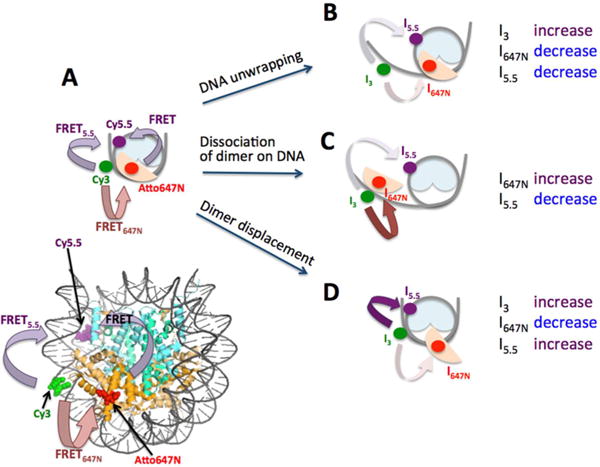
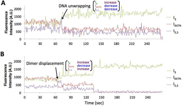
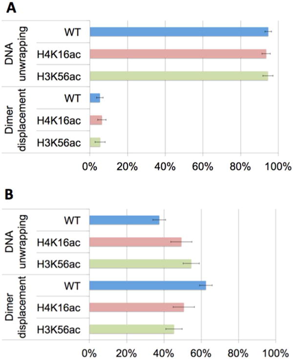
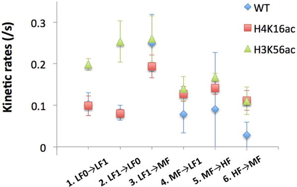
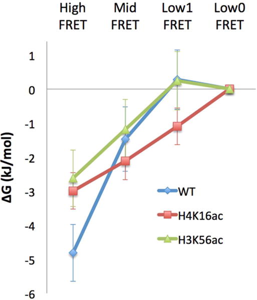
Nucleosomes impose physical barriers to DNA-templated processes, playing important roles in eukaryotic gene regulation. DNA is packaged into nucleosomes by histone proteins mainly through strong electrostatic interactions that can be modulated by various post-translational histone modifications. Investigating the dynamics of histone dissociation from the nucleosome and how it is altered upon histone modifications is important for understanding eukaryotic gene regulation mechanisms. In particular, histone H2A-H2B dimer displacement in the nucleosome is one of the most important and earliest steps of histone dissociation. Two conflicting hypotheses on the requirement for dimer displacement are that nucleosomal DNA needs to be unwrapped before a dimer can displace and that a dimer can displace without DNA unwrapping. In order to test the hypotheses, we employed three-color single-molecule FRET and monitored in a time-resolved manner the early kinetics of H2A-H2B dimer dissociation triggered by high salt concentration and by histone chaperone Nap1. The results reveal that dimer displacement requires DNA unwrapping in the vast majority of the nucleosomes in the salt-induced case, while dimer displacement precedes DNA unwrapping in >60% of the nucleosomes in the Nap1-mediated case. We also found that acetylation at histone H4K16 or H3K56 affects the kinetics of Nap1-mediated dimer dissociation and facilitates the process both kinetically and thermodynamically. On the basis of these results, we suggest a mechanism by which histone chaperone facilitates H2A-H2B dimer displacement from the histone core without requiring another factor to unwrap the nucleosomal DNA.


Conflict of interest statement

Notes

The authors declare no competing financial interest.

Figures

Figure 1
Figure 1
Three-color FRET experimental setup to monitor the early steps of histone H2A-H2B dimer dissociation. (A) A schematic representation of a complete nucleosome core particle with intact DNA and H2A-H2B dimer reporting moderately high FRET5.5 and FRET647N. Fluorescence intensities from Cy3, Atto647N, and Cy5.5 are denoted by I3, I647N, and I5.5, respectively. (B) A nucleosome with unwrapped DNA and intact dimer would report decreasing I5.5 and I647N and increasing I3. (C) A nucleosome with DNA and dimer dissociating simultaneously would show decreasing I5.5 and increasing I647N. This mode of dissociation can be via gyre opening or DNA unwrapping, which cannot be distinguished in our setup. (D) A nucleosome with displaced dimer with intact DNA would report increasing I5.5 and I3, and decreasing I647N.
Figure 2
Figure 2
Representative time traces of fluorescence intensities during the early steps of dimer dissociation (A.U. = arbitrary unit). More traces are shown in Figure S4. (A) DNA unwrapping initiates dimer dissociation as characterized by simultaneously decreasing I5.5 and I647N with increasing I3. This trace is from the wild type nucleosome under the high salt condition. The acceptor fluocesnce signals are eventually extingiuished at the end of the time trace by either dimer dissociation or photobleaching within 1−2 frames at ~270 s. (B) Dimer displacement initiates dissociation as characterized by increasing I5.5 and simultaneously decreasing I647N and increasing I3. This trace is from the wild type nucleosome under the Nap1 condition. The increased donor intensity and the decreased acceptor intensities at 135 s are due to either Atto647N photobleaching or dimer dissociation.
Figure 3
Figure 3
Population densities of nucleosomes grouped by the initial dimer dissociation step. The two populations are denoted by “DNA unwrapping” and “Dimer displacement”. Nucleosomes in the “DNA unwrapping” group start dimer dissociation by DNA unwrapping prior to dimer displacement. Nucleosomes in the “Dimer displacement” group start dimer dissociation by dimer displacement prior to DNA unwrapping. Dimer displacement was induced by (A) a high salt level and (B) Nap1. WT, H4K16ac, and H3K56ac denote wild-type nucleosome (i.e., no acetylation), nucleosome acetylated at H4K16, and nucleosome acetylated at H3K56, respectively. The error bars represent the standard deviations of the counts assuming a binomial distribution whose variance is given by np(1 − p), where n is the sample size and p is the probability. (A) In the 95% of the population, dimer dissociation induced by salt starts with DNA unwrapping and the effect of acetylation on this statistics is negligible. (B) In the 63% of the population, dimer dissociation mediated by Nap1 starts with dimer displacement (wild-type, WT). This population density decreases significantly upon histone acetylation at H4K16 or H3K56 (Table 1C).
Figure 4
Figure 4
Kinetic rates between two adjacent states in the four FRET states analyzed by hidden Markov models. WT denotes wild-type, or no acetylation. Acetylations at H4K16 (H4K16ac) and H3K56 (H3K56ac) increase the kinetic rate from High to Mid FRET. H3K56 acetylation facilitates the DNA fluctuations between the Low1 and Low0 FRET states. The errors are the standard deviations obtained from five HMM analyses per case (see Materials and Methods). The rates and errors are listed in Table 3.
Figure 5
Figure 5
Relative thermodynamic stabilities of the early intermediates of dimer dissociation. WT, H4K16ac, and H3K56ac denote wild-type nucleosome (i.e., no acetylation), nucleosome acetylated at H4K16, and nucleosome acetylated at H3K56, respectively. The relative free energies (ΔG) were derived from the equilibrium population densities obtained by numerical simulations based on the kinetic rates listed in Table 3. The free energy values were aligned to the Low0 FRET state assuming that this state has a thermodynamic stability unaffected by the acetylation state. The error bars represent the standard deviations of five simulations based on five different sets of kinetic rates estimated from five different sets of HMM analysis results (see Materials and Methods). The ΔG values are listed in Table 4. RT at 298 K is 2.48 kJ/mol.

Similar articles

Cited by

References

    1. Luger K, Mader AW, Richmond RK, Sargent DF, Richmond TJ. Crystal structure of the nucleosome core particle at 2.8 Å resolution. Nature. 1997;389:251–260. - PubMed
    1. Hoch DA, Stratton JJ, Gloss LM. Protein-protein Forster resonance energy transfer analysis of nucleosome core particles containing H2A and H2A.Z. J Mol Biol. 2007;371:971–988. - PMC - PubMed
    1. Yager TD, McMurray CT, van Holde KE. Salt-induced release of DNA from nucleosome core particles. Biochemistry. 1989;28:2271–2281. - PubMed
    1. Okuwaki M, Kato K, Shimahara H, Tate S, Nagata K. Assembly and disassembly of nucleosome core particles containing histone variants by human nucleosome assembly protein I. Mol Cell Biol. 2005;25:10639–10651. - PMC - PubMed
    1. Andrews AJ, Chen X, Zevin A, Stargell LA, Luger K. The histone chaperone Nap1 promotes nucleosome assembly by eliminating nonnucleosomal histone DNA interactions. Mol Cell. 2010;37:834–842. - PMC - PubMed

Publication types

MeSH terms

LinkOut - more resources