

"Black Bone" MRI: a novel imaging technique for 3D printing
- PMID: 28128636
- PMCID: PMC5606274
- DOI: 10.1259/dmfr.20160407
"Black Bone" MRI: a novel imaging technique for 3D printing
Abstract
Objectives: Three-dimensionally printed anatomical models are rapidly becoming an integral part of pre-operative planning of complex surgical cases. We have previously reported the "Black Bone" MRI technique as a non-ionizing alternative to CT. Segmentation of bone becomes possible by minimizing soft tissue contrast to enhance the bone-soft tissue boundary. The objectives of this study were to ascertain the potential of utilizing this technique to produce three-dimensional (3D) printed models.
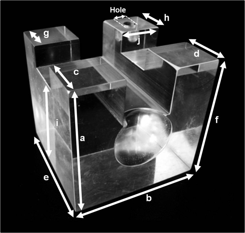
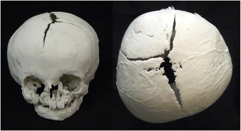
Methods: "Black Bone" MRI acquired from adult volunteers and infants with craniosynostosis were 3D rendered and 3D printed. A custom phantom provided a surrogate marker of accuracy permitting comparison between direct measurements and 3D printed models created by segmenting both CT and "Black Bone" MRI data sets using two different software packages.
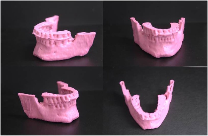
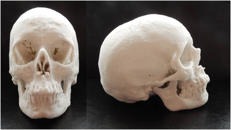
Results: "Black Bone" MRI was successfully utilized to produce 3D models of the craniofacial skeleton in both adults and an infant. Measurements of the cube phantom and 3D printed models demonstrated submillimetre discrepancy.
Conclusions: In this novel preliminary study exploring the potential of 3D printing from "Black Bone" MRI data, the feasibility of producing anatomical 3D models has been demonstrated, thus offering a potential non-ionizing alterative to CT for the craniofacial skeleton.
Keywords: 3D imaging; 3D printing; MRI; anatomical models; segmentation.
Figures
References
-
- Bibb R. Pre-operative management—modelling and rapid prototyping. Imaging and planning in surgery: a guide to research. Switzerland: AO Publishing; 2008.
-
- Eley KA, Richards R, Dobson D, Linney A, Watt-Smith SR. Re.: Development of in-house rapid manufacturing of three-dimensional models in maxillofacial surgery. Br J Oral Maxillofac Surg 2011; 49: 326–7. doi: https://doi.org/10.1016/j.bjoms.2010.09.019 - DOI - PubMed
-
- Lill W, Solar P, Ulm C, Watzek G, Blahout R, Matejka M. Reproducibility of three-dimensional CT-assisted model production in the maxillofacial area. Br J Oral Maxillofac Surg 1992; 30: 233–6. doi: https://doi.org/10.1016/0266-4356(92)90265-K - DOI - PubMed
-
- Sailer HF, Haers PE, Zollikofer CP, Warnke T, Carls FR, Stucki P. The value of stereolithographic models for preoperative diagnosis of craniofacial deformities and planning of surgical corrections. Int J Oral Maxillofac Surg 1998; 27: 327–33. doi: https://doi.org/10.1016/S0901-5027(98)80059-3 - DOI - PubMed
-
- Imai K, Tsujiguchi K, Toda C, Enoki E, Sung KC, Sakamoto H, et al. . Reduction of operating time and blood transfusion for craniosynostosis by simulated surgery using three-dimensional solid models. Neurol Med Chir (Tokyo) 1999; 39: 423–6; discussion 427. doi: https://doi.org/10.2176/nmc.39.423 - DOI - PubMed
MeSH terms
LinkOut - more resources
Full Text Sources
Other Literature Sources
Medical
