. 2017 Jan 4;25(1):12-23.
doi: 10.1016/j.ymthe.2016.11.010. Epub 2017 Jan 4.

CRISPR/Cas9 Editing of the Mutant Huntingtin Allele In Vitro and In Vivo

Affiliations

CRISPR/Cas9 Editing of the Mutant Huntingtin Allele In Vitro and In Vivo

Alex Mas Monteys et al. Mol Ther. .

Abstract

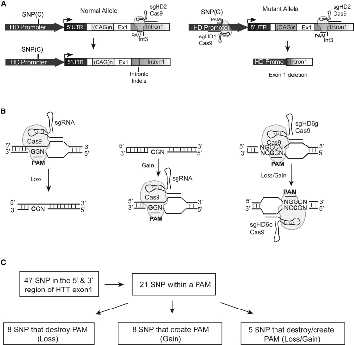
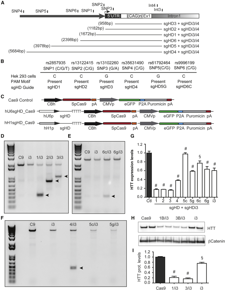
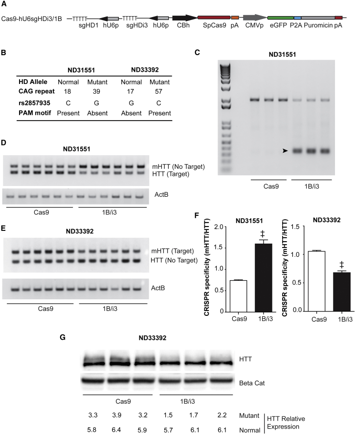
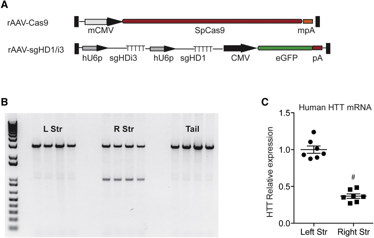
Huntington disease (HD) is a fatal dominantly inherited neurodegenerative disorder caused by CAG repeat expansion (>36 repeats) within the first exon of the huntingtin gene. Although mutant huntingtin (mHTT) is ubiquitously expressed, the brain shows robust and early degeneration. Current RNA interference-based approaches for lowering mHTT expression have been efficacious in mouse models, but basal mutant protein levels are still detected. To fully mitigate expression from the mutant allele, we hypothesize that allele-specific genome editing can occur via prevalent promoter-resident SNPs in heterozygosity with the mutant allele. Here, we identified SNPs that either cause or destroy PAM motifs critical for CRISPR-selective editing of one allele versus the other in cells from HD patients and in a transgenic HD model harboring the human allele.

Keywords: AAV; CRISPR/Cas9; Huntington’s disease; gene therapy; genome editing.


Figures

Figure 1
Figure 1
SNP-Dependent Editing for Huntington Disease Therapy (A) Cartoon depicting the allele-specific editing strategy to abrogate mutant HTT expression. SNPs within PAM sequences upstream of HTT exon-1 permit specific targeted deletions of the mutant allele when present in heterozygosity. After DNA repair, mutant HTT exon-1 is deleted by a pair of sgRNA/Cas9 complexes binding upstream and downstream of exon-1 (right), whereas intronic indels could be generated by a single dsDNA break in the normal allele (left). (B) The nucleotide variation of a SNP within a PAM alters Cas9 recognition resulting in the loss (left), the gain (middle), or the simultaneous loss of a PAM in one DNA strand and the gain of a PAM on the opposite strand (right). (C) There are 21 out of 47 prevalent SNPs flanking HTT exon-1 that are located within predicted critical positions of a PAM sequence for the CRISPR/SpCas9 system analyzed. The minor frequency allele either mediates the loss (eight SNPs), gain (eight SNPs), or a loss/gain (five SNPs) of a PAM motif.
Figure 2
Figure 2
Cleavage of SNP-Dependent sgHD/SpCas9 Complexes in HEK293 Cells (A) Cartoon depicting the relative position of the six prevalent SNP-dependent PAMs upstream of HTT exon-1 and two common PAMs within HTT intron-1. The estimated size of the targeted deleted sequence is indicated. (B) The genotype of the prevalent SNPs within the HTT promoter in HEK293 cells is shown. All SNPs were homozygous for the nucleotide variation and the PAM motif was present for the sgRNA indicated. (C) A diagram of the CRISPR expression systems transfected into HEK293 cells is shown. (D–F) A genomic PCR showing HTT exon-1-targeted deletion by sgRNA/SpCas9 pair complexes binding upstream and downstream of the target sequence is shown in the images. (G) RT-qPCR analysis of HTT mRNA levels in HEK293 cells transfected with sgHD/SpCas9 expression cassettes targeting upstream promoter SNPs and the common intronic sgHDi3 sequence is shown. All of the samples are normalized to human GAPDH, and the results are the mean ± SEM relative to cells transfected with plasmids containing the SpCas9 only control (n = 6 independent experiments; §p < 0.001, #p < 0.0001, and one-way ANOVA followed by a Bonferroni’s post hoc). (H) sgHD1/i3/SpCas9, sgHD3/i3/SpCas9, and sgHDi3/SpCas9 expression cassettes were transfected into HEK293 cells, and endogenous HTT protein levels were determined after puromycin selection and expansion. Cells transfected with Cas9 only were used as a control and beta catenin served as a loading control. (I) The quantification of HTT protein levels after treatment with sgHD/SpCas9 complexes is shown. The data are the mean ± SEM relative to cells transfected with plasmids containing SpCas9 only control (n = 6 independent experiments; #p < 0.0001, §p < 0.001, and one-way ANOVA followed by Bonferroni’s post hoc).
Figure 3
Figure 3
Assessment of Allele-Specific Cleavage in Human HD Fibroblasts (A) Cartoon depicting the CRISPR expression plasmid used to co-express sgHD1 and sgHDi3 expression cassettes. SpCas9 and the selective reporter eGFP/puromycin expression cassettes present in the same plasmid are also shown. (B) ND31551 and ND33392 HD fibroblasts lines were selected to determine allele-specific deletion of HTT. CAG repeat length, nucleotide variation, and the allele location of the PAM motif are indicated in the image. (C) A representative genomic PCR showing HTT exon-1 deletion of DNA harvested from the electroporated ND31551 HD fibroblast cell line is shown in the image. The arrow indicates the expected PCR amplification product resulting from allele-specific deletion. (D and E) A semiquantitative PCR reaction showing the reduction of the targeted allele containing the conserved PAM sequence is shown in the image. For ND31551 fibroblasts, the PAM sequence is conserved in the normal allele, while for ND33392 fibroblasts, the PAM sequence is in the mutant allele. The expression levels are reduced only on the PAM-containing allele. (F) The quantification of mRNA reduction in treated HD fibroblasts is shown. The data show the ratio between mRNA levels of the mutant with respect to the normal allele, relative to cells electroporated with vectors expressing only the Cas9 control. The results are mean ± SEM relative to cells transfected with plasmids containing SpCas9 only control (n = 6; individual electroporations per group, ‡p < 0.01, Mann-Whitney t test). (G) A representative western blot showing allele-specific depletion (upper band of HTT doublet consisting of normal [lower] and expanded polyQ-containing proteins) after electroporation of HD fibroblasts with sgHD/SpCas9 expression vectors is shown in the image. The numbers below the image refer to relative HTT levels.
Figure 4
Figure 4
Assessing Off-Target Activity of sgHD1 and sgHDi3/Cas9 (A) Table depicting the number of off-target sites for the most active sequences predicted to bind with 1, 2, or 3 mismatches. The nucleotide length of the complementary guide sequence is also indicated in the table. (B) Table highlighting the number of off-target sites binding at different genomic regions using the UCSC genome browser is shown. (C) The HD fibroblasts were electroporated with plasmids expressing sgHD1/i3 and SpCas9 along with an ODN sequence. The Sanger sequencing results showed the incorporation of the ODN sequence at the DNA cleavage site. A HTT promoter sequence and a HTT intron sequence outside the ODN sequence are also depicted. (D) Sanger sequencing results from 11 predicted off-target sites are shown. The gene name, chromosome position, DNA strand, number of mismatches and position within the guide, gene location, sgRNA sequence, and indel presence or absence are indicated.
Figure 5
Figure 5
In Vivo Gene Editing of the Mutant HTT Allele (A) Cartoon depicting rAAV shuttle vectors containing the SpCas9 and sgHD1/i3 expression cassettes (mCMV, minimal CMV promoter; mpA, minimal polyA and hU6p, human U6 promoter; pA, SV40 polyA). (B) PCR of isolated genomic DNA showing human HTT exon-1 targeted deletion after injection of vectors expressing SpCas9 and sgRNA sequences is shown. left striatum, LStr; right striatum, RStr. (C) qRT-PCR analysis of HTT mRNA levels in striatum samples harvested 3 weeks after SpCas9 and sgHD1/i3 delivery is shown. All of the samples were normalized to beta actin and the results are mean ± SEM relative to uninjected striatal samples (n = 7 animals per group, #p < 0.0001, and unpaired t test).

Similar articles

Cited by

References

    1. Walker F.O. Huntington’s disease. Semin. Neurol. 2007;27:143–150. - PubMed
    1. Hicks R.R., Smith D.H., Lowenstein D.H., Saint Marie R., McIntosh T.K. Mild experimental brain injury in the rat induces cognitive deficits associated with regional neuronal loss in the hippocampus. J. Neurotrauma. 1993;10:405–414. - PubMed
    1. Johnson C.D., Davidson B.L. Huntington’s disease: progress toward effective disease-modifying treatments and a cure. Hum. Mol. Genet. 2010;19(R1):R98–R102. - PMC - PubMed
    1. Yamamoto A., Lucas J.J., Hen R. Reversal of neuropathology and motor dysfunction in a conditional model of Huntington’s disease. Cell. 2000;101:57–66. - PubMed
    1. Díaz-Hernández M., Torres-Peraza J., Salvatori-Abarca A., Morán M.A., Gómez-Ramos P., Alberch J., Lucas J.J. Full motor recovery despite striatal neuron loss and formation of irreversible amyloid-like inclusions in a conditional mouse model of Huntington’s disease. J. Neurosci. 2005;25:9773–9781. - PMC - PubMed

MeSH terms