. 2017 Jan 4;25(1):205-217.
doi: 10.1016/j.ymthe.2016.10.015. Epub 2017 Jan 4.

HOTAIR Epigenetically Modulates PTEN Expression via MicroRNA-29b: A Novel Mechanism in Regulation of Liver Fibrosis

Affiliations

HOTAIR Epigenetically Modulates PTEN Expression via MicroRNA-29b: A Novel Mechanism in Regulation of Liver Fibrosis

Fujun Yu et al. Mol Ther. .

Erratum in

Abstract

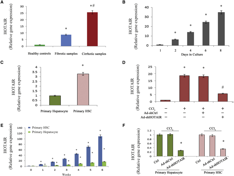
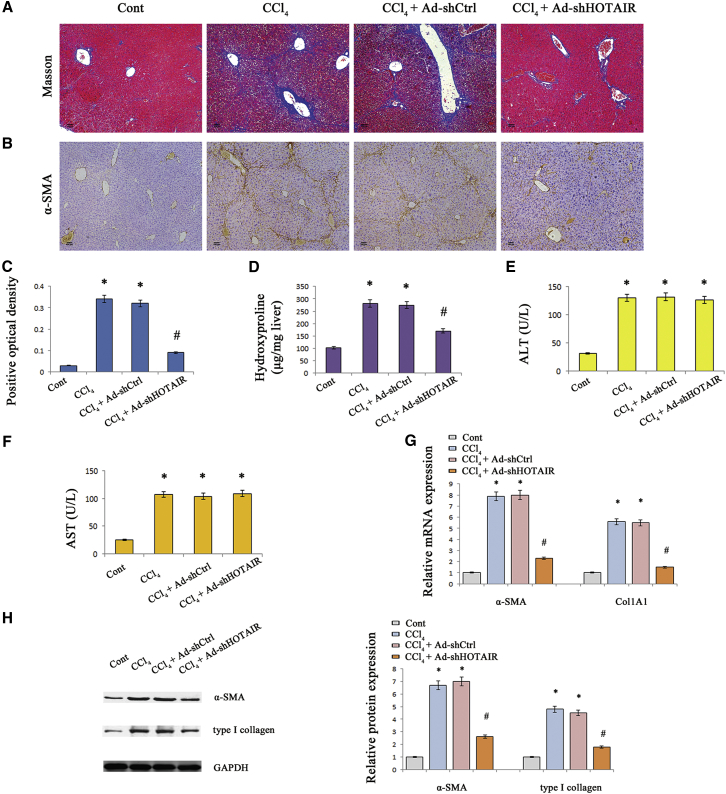
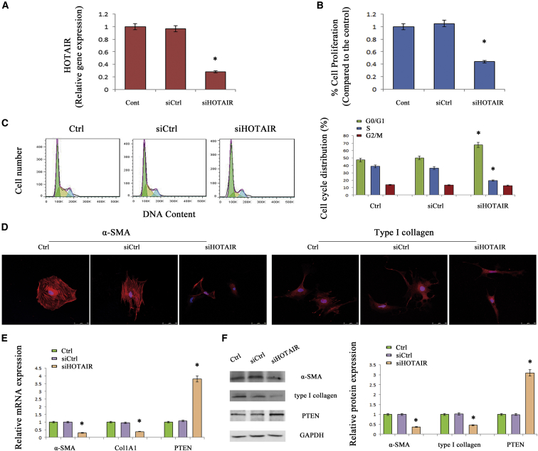
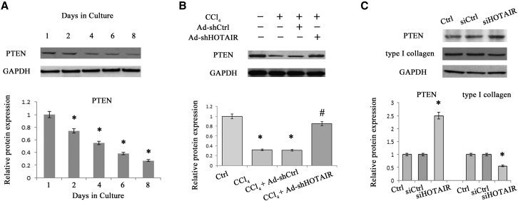
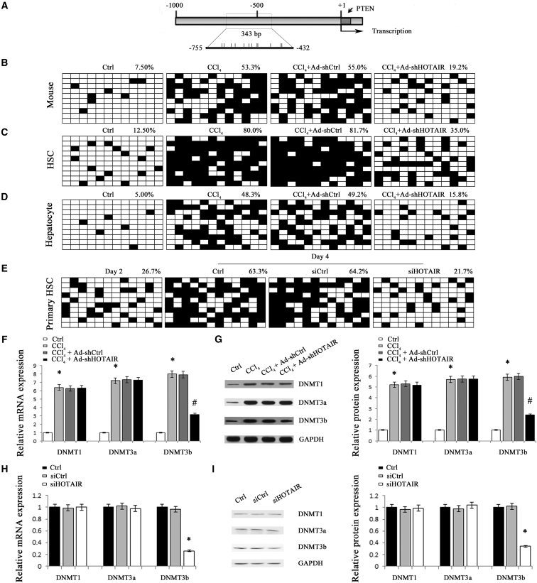
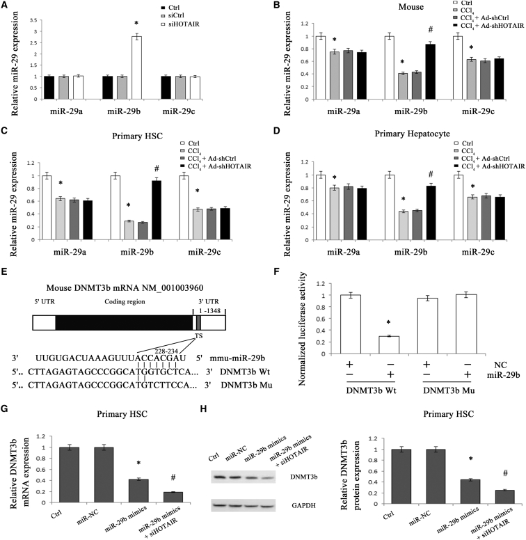
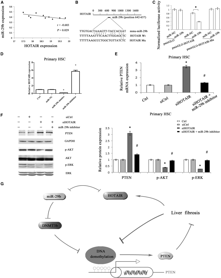
Homeobox transcript antisense RNA (HOTAIR), as a long intergenic non-coding RNA (lincRNA), is upregulated in various cancers and involved in diverse cellular functions. However, its role in liver fibrosis is unclear. In this study, HOTAIR expression was upregulated in hepatic stellate cells (HSCs) in vivo and in vitro during liver fibrosis. HOTAIR knockdown suppressed HSC activation including α-smooth muscle actin (α-SMA) and typeIcollagen in vitro and in vivo. Both HSC proliferation and cell cycle were inhibited by HOTAIR knockdown. Notably, inhibition of HOTAIR led to an increase in PTEN, associated with the loss of DNA methylation. miR-29b-mediated control of PTEN methylation was involved in the effects of HOTAIR knockdown. HOTAIR was confirmed a target of miR-29b and lack of the miR-29b binding site in HOTAIR prevented the suppression of miR-29b, suggesting HOTAIR contributes to PTEN expression downregulation via sponging miR-29b. Interestingly, increased HOTAIR was also observed in hepatocytes during liver fibrosis. Loss of HOTAIR additionally led to the increase in PTEN and the reduction in typeIcollagen in hepatocytes. Collectively, we demonstrate that HOTAIR downregulates miR-29b expression and attenuates its control on epigenetic regulation, leading to enhanced PTEN methylation, which contributes to the progression of liver fibrosis.

Keywords: DNA methylation; DNA methyltransferase; DNMT; HOTAIR; PTEN; homeobox transcript antisense RNA; microRNA-29b; phosphatase and tensin homolog deleted on chromosome 10.


Figures

Figure 1
Figure 1
HOTAIR Is Upregulated during Liver Fibrosis (A) HOTAIR was increased in liver cirrhotic samples (n = 15) and fibrotic samples (n = 15) when compared to healthy controls (n = 15). (B) Relative HOTAIR gene expression was detected in primary HSCs during culture days. (C) HOTAIR was detected in primary hepatocytes and primary HSCs from the livers of healthy mice. (D) HOTAIR was analyzed in the liver from CCl4 mice after Ad-shHOTAIR treatment. (E) HOTAIR was detected in isolated primary HSCs and primary hepatocytes from CCl4 mice at different weeks. (F) HOTAIR was detected in isolated primary hepatocytes and primary HSCs from CCl4 mice after Ad-shHOTAIR treatment. *p < 0.05 compared to the control and #p < 0.05 compared to fibrotic samples or CCl4 group. Each value is the mean ± SD of three experiments.
Figure 2
Figure 2
HOTAIR Downregulation Suppressed CCl4-Induced Liver Fibrosis in Mice (A) Accumulation of collagen was assessed by Masson staining. The scale bar represents 100 μm. (B) α-SMA level was analyzed by immunohistochemistry. The scale bar represents 100 μm. The levels of α-SMA positive optical density (C) and hydroxyproline (D) were analyzed in CCl4 mice after Ad-shHOTAIR treatment. The levels of ALT (E) and AST (F) were analyzed after Ad-shHOTAIR treatment. The mRNA (G) and protein (H) expressions of α-SMA and Col1A1 were analyzed after Ad-shHOTAIR treatment. *p < 0.05 compared to the control and #p < 0.05 compared to CCl4 group. Each value is the mean ± SD of three experiments.
Figure 3
Figure 3
Effects of siHOTAIR on HSC Activation HSCs were transfected with siHOTAIR for 48 hr. The effects of siHOTAIR on HOTAIR level (A), HSC proliferation (B), and cell cycle (C) are shown. HSC proliferation was detected by MTT assay. (D) Immunofluorescence staining for α-SMA (red) and type I collagen (red) were evaluated by confocal laser microscopy. DAPI stained the nuclei blue. The scale bar represents 50 μm. The mRNA (E) and protein (F) expressions of α-SMA, Col1A1, and PTEN were analyzed. *p < 0.05 compared to the control and each value is the mean ± SD of three experiments.
Figure 4
Figure 4
Effects of siHOTAIR on PTEN Expression (A) PTEN protein expression was detected in primary HSCs. (B) PTEN protein expression was detected in CCl4 mice after Ad-shHOTAIR. (C) PTEN protein and type I collagen were analyzed in primary hepatocytes transfected with siHOTAIR. *p < 0.05 compared to the control and #p < 0.05 compared to CCl4 group. Each value is the mean ± SD of three experiments.
Figure 5
Figure 5
PTEN Expression Was Regulated by Promoter DNA Methylation CCl4 mice were treated with Ad-shHOTAIR. (A) A schematic representation of the promoter region amplified by bisulfide sequencing. Each vertical bar represents the presence of a CpG dinucleotide. The average percentage of PTEN methylation was shown in the livers from mice (B), as well as isolated primary HSCs (C), and primary hepatocytes (D). (E) The average percentage of PTEN methylation was shown in HSCs with siHOTAIR. The mRNA (F) and protein (G) expression levels of DNMT1, DNMT3a, and DNMT3b were analyzed in CCl4 mice after Ad-shHOTAIR treatment. The mRNA (H) and protein (I) expression levels of DNMT1, DNMT3a, and DNMT3b were analyzed in primary HSCs transfected with siHOTAIR. *p < 0.05 compared to the control and #p < 0.05 compared to CCl4 group. Each value is the mean ± SD of three experiments.
Figure 6
Figure 6
Silencing HOTAIR Enhanced miR-29b Expression (A) The levels of miR-29a, miR-29b, and miR-29c were analyzed in HSCs transfected with siHOTAIR. CCl4 mice were treated with Ad-shHOTAIR. The levels of miR-29a, miR-29b, and miR-29c were detected in the livers from mice (B), as well as isolated primary HSCs (C), and primary hepatocytes (D). (E) Putative miR-29b binding sites (TS) within the mouse DNMT3b 3′-UTR are shown. The position of the binding sites was numbered relative to the first nucleotide of the 3′-UTR. Mutations were introduced into DNMT3b 3′-UTR that matched the seed region of miR-29b as shown in DNMT3b Mu. (F) Dual-luciferase assay was performed in HEK293T co-transfected with luciferase constructs containing the DNMT3b wild-type or Mu 3′-UTR and miR-29b mimics or scrambled oligonucleotides as the negative control. The mRNA (G) and protein (H) levels of DNMT3b in primary HSCs were reduced by miR-29b mimics, which were further decreased by siHOTAIR. *p < 0.05 compared to the control and #p < 0.05 compared to CCl4 or miR-29b mimics group. Each value is the mean ± SD of three experiments.
Figure 7
Figure 7
The Effect of HOTAIR on PTEN Expression Is through Competitively Binding miR-29b (A) Correlation between HOTAIR level and miR-29b expression in liver fibrosis tissue samples from CCl4 mice (n = 10) was subjected to Pearson correlation analysis. (B) Schematic diagram of the miR-29b binding site in the HOTAIR based on RNA22 software. (C) Relative luciferase activities of luciferase reporters bearing wild-type or mutant HOTAIR were analyzed 48 hr following transfection with the indicated miR-29b mimics or miR-NC in HEK293T. Relative gene expressions of HOTAIR (D) and PTEN (E) were analyzed by quantitative real-time PCR. (F) PTEN, phosphorylation of ERK (T202/Y204) and Akt (S473) were analyzed by western blotting. (G) The signal pathway was discovered in liver fibrosis. *p < 0.05 compared to the control and #p < 0.05 compared to siHOTAIR group. Each value is the mean ± SD of three experiments.

References

    1. Kong X., Horiguchi N., Mori M., Gao B. Cytokines and STATs in Liver Fibrosis. Front. Physiol. 2012;3:69. - PMC - PubMed
    1. Kwiecinski M., Noetel A., Elfimova N., Trebicka J., Schievenbusch S., Strack I., Molnar L., von Brandenstein M., Töx U., Nischt R. Hepatocyte growth factor (HGF) inhibits collagen I and IV synthesis in hepatic stellate cells by miRNA-29 induction. PLoS ONE. 2011;6:e24568. - PMC - PubMed
    1. Wang J., Chu E.S., Chen H.Y., Man K., Go M.Y., Huang X.R., Lan H.Y., Sung J.J., Yu J. microRNA-29b prevents liver fibrosis by attenuating hepatic stellate cell activation and inducing apoptosis through targeting PI3K/AKT pathway. Oncotarget. 2015;6:7325–7338. - PMC - PubMed
    1. Friedman S.L. Hepatic stellate cells: protean, multifunctional, and enigmatic cells of the liver. Physiol. Rev. 2008;88:125–172. - PMC - PubMed
    1. Schickel R., Boyerinas B., Park S.M., Peter M.E. MicroRNAs: key players in the immune system, differentiation, tumorigenesis and cell death. Oncogene. 2008;27:5959–5974. - PubMed

MeSH terms

Substances