

Object crowding in age-related macular degeneration
- PMID: 28129416
- PMCID: PMC5283087
- DOI: 10.1167/17.1.33
Object crowding in age-related macular degeneration
Abstract
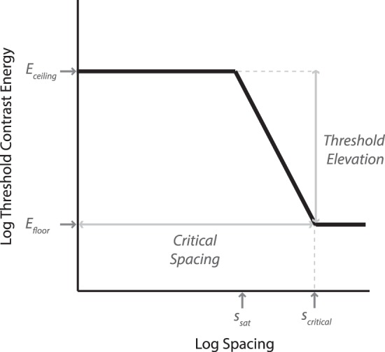
Crowding, the phenomenon of impeded object identification due to clutter, is believed to be a key limiting factor of form vision in the peripheral visual field. The present study provides a characterization of object crowding in age-related macular degeneration (AMD) measured at the participants' respective preferred retinal loci with binocular viewing. Crowding was also measured in young and age-matched controls at the same retinal locations, using a fixation-contingent display paradigm to allow unlimited stimulus duration. With objects, the critical spacing of crowding for AMD participants was not substantially different from controls. However, baseline contrast energy thresholds in the noncrowded condition were four times that of the controls. Crowding further exacerbated deficits in contrast sensitivity to three times the normal crowding-induced contrast energy threshold elevation. These findings indicate that contrast-sensitivity deficit is a major limiting factor of object recognition for individuals with AMD, in addition to crowding. Focusing on this more tractable deficit of AMD may lead to more effective remediation and technological assistance.
Figures


Similar articles
-
Mesopic Pelli-Robson contrast sensitivity and MP-1 microperimetry in healthy ageing and age-related macular degeneration.Acta Ophthalmol. 2016 Dec;94(8):e772-e778. doi: 10.1111/aos.13112. Epub 2016 May 26. Acta Ophthalmol. 2016. PMID: 27225020
-
Object crowding.J Vis. 2011 May 25;11(6):10.1167/11.6.19 19. doi: 10.1167/11.6.19. J Vis. 2011. PMID: 21613388 Free PMC article.
-
Scene perception in age-related macular degeneration.Invest Ophthalmol Vis Sci. 2010 Dec;51(12):6868-74. doi: 10.1167/iovs.10-5517. Invest Ophthalmol Vis Sci. 2010. PMID: 21123770
-
Determinants of concern about falling in adults with age-related macular degeneration.Ophthalmic Physiol Opt. 2021 Mar;41(2):245-254. doi: 10.1111/opo.12777. Epub 2020 Dec 25. Ophthalmic Physiol Opt. 2021. PMID: 33368495
-
Binocular function in patients with age-related macular degeneration: a review.Can J Ophthalmol. 2006 Jun;41(3):327-32. doi: 10.1139/I06-029. Can J Ophthalmol. 2006. PMID: 16767188 Review.
Cited by
-
Anodal transcranial direct current stimulation reduces collinear lateral inhibition in normal peripheral vision.PLoS One. 2020 May 6;15(5):e0232276. doi: 10.1371/journal.pone.0232276. eCollection 2020. PLoS One. 2020. PMID: 32374787 Free PMC article.
-
Eye Movements in Macular Degeneration.Annu Rev Vis Sci. 2021 Sep 15;7:773-791. doi: 10.1146/annurev-vision-100119-125555. Epub 2021 May 26. Annu Rev Vis Sci. 2021. PMID: 34038144 Free PMC article. Review.
-
Mixture model investigation of the inner-outer asymmetry in visual crowding reveals a heavier weight towards the visual periphery.Sci Rep. 2021 Jan 22;11(1):2116. doi: 10.1038/s41598-021-81533-9. Sci Rep. 2021. PMID: 33483608 Free PMC article.
-
An exploratory study to evaluate visual function endpoints in non-advanced age-related macular degeneration.BMC Ophthalmol. 2020 Oct 22;20(1):424. doi: 10.1186/s12886-020-01683-8. BMC Ophthalmol. 2020. PMID: 33092549 Free PMC article.
-
Is the Normal Periphery in Young Adults a Good Model for Reading in the Presence of Central Vision Loss?Invest Ophthalmol Vis Sci. 2025 Feb 3;66(2):62. doi: 10.1167/iovs.66.2.62. Invest Ophthalmol Vis Sci. 2025. PMID: 39992670 Free PMC article.
References
-
- Andriessen, J. J.,& Bouma, H.. (1976). Eccentric vision: Adverse interactions between line segments. Vision Research, 16 1, 71–78. - PubMed
-
- Bouma, H. (1970, Apr 1). Interaction effects in parafoveal letter recognition. Nature, 226 5241, 177–178. - PubMed
-
- Bouma, H. (1973). Visual interference in the parafoveal recognition of initial and final letters of words. Vision Research, 13 4, 767–782. - PubMed
-
- Calabrèse, A., Bernard, J. B., Hoffart, L., Faure, G., Barouch, F., Conrath, J.,& Castet, E.. (2010). Small effect of interline spacing on maximal reading speed in low-vision patients with central field loss irrespective of scotoma size. Investigative Ophthalmology and Visual Science, 51, 1247–1254. [PubMed] [Article] - PubMed
-
- Choudhury, A.,& Medioni, G.. (2010). Color contrast enhancement for visually impaired people. 2010 IEEE Computer Society Conference on Computer Vision and Pattern Recognition - Workshops (pp 33–40. New York: IEEE.
MeSH terms
Grants and funding
LinkOut - more resources
Full Text Sources
Other Literature Sources
Medical
