. 2017 Jan 28;8(1):19.
doi: 10.1186/s13287-017-0475-8.

Human umbilical cord-derived mesenchymal stromal cells protect against premature renal senescence resulting from oxidative stress in rats with acute kidney injury

Affiliations

Human umbilical cord-derived mesenchymal stromal cells protect against premature renal senescence resulting from oxidative stress in rats with acute kidney injury

Camila Eleuterio Rodrigues et al. Stem Cell Res Ther. .

Abstract

Background: Mesenchymal stromal cells (MSCs) represent an option for the treatment of acute kidney injury (AKI). It is known that young stem cells are better than are aged stem cells at reducing the incidence of the senescent phenotype in the kidneys. The objective of this study was to determine whether AKI leads to premature, stress-induced senescence, as well as whether human umbilical cord-derived MSCs (huMSCs) can prevent ischaemia/reperfusion injury (IRI)-induced renal senescence in rats.

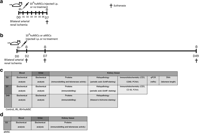
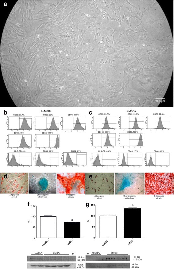
Methods: By clamping both renal arteries for 45 min, we induced IRI in male rats. Six hours later, some rats received 1 × 10⁶ huMSCs or human adipose-derived MSCs (aMSCs) intraperitoneally. Rats were euthanised and studied on post-IRI days 2, 7 and 49.

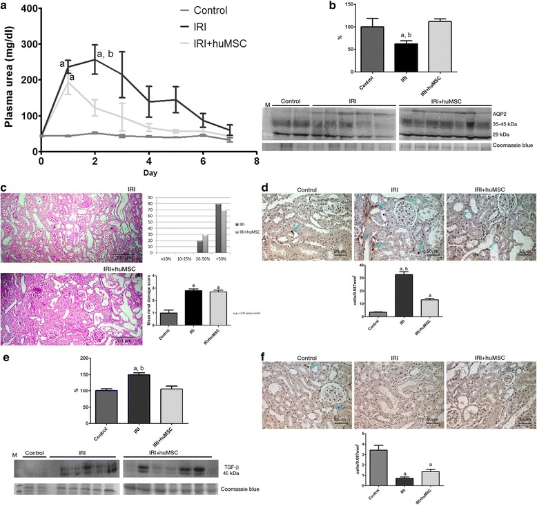
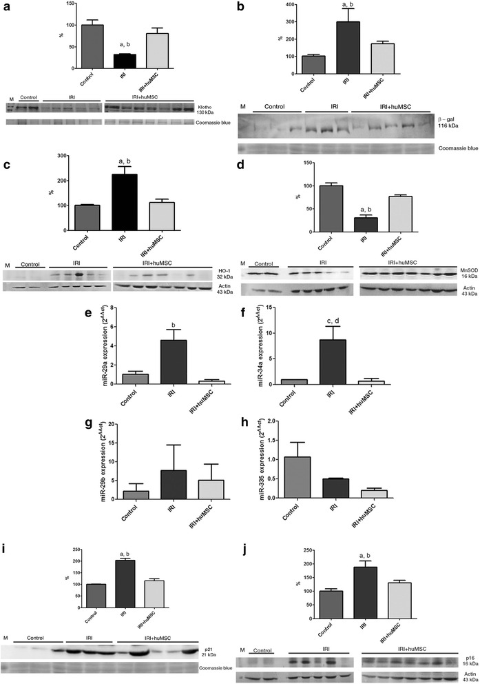
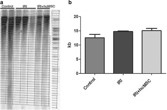
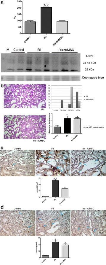
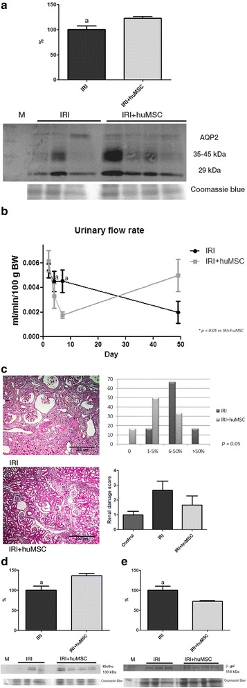
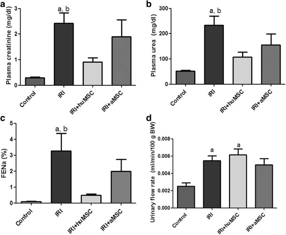
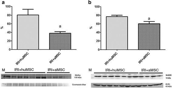
Results: On post-IRI day 2, the kidneys of huMSC-treated rats showed improved glomerular filtration, better tubular function and higher expression of aquaporin 2, as well as less macrophage infiltration. Senescence-related proteins (β-galactosidase, p21Waf1/Cip1, p16INK4a and transforming growth factor beta 1) and microRNAs (miR-29a and miR-34a) were overexpressed after IRI and subsequently downregulated by the treatment. The IRI-induced pro-oxidative state and reduction in Klotho expression were both reversed by the treatment. In comparison with huMSC treatment, the treatment with aMSCs improved renal function to a lesser degree, as well as resulting in a less pronounced increase in the renal expression of Klotho and manganese superoxide dismutase. Treatment with huMSCs ameliorated long-term kidney function after IRI, minimised renal fibrosis, decreased β-galactosidase expression and increased the expression of Klotho.

Conclusions: Our data demonstrate that huMSCs attenuate the inflammatory and oxidative stress responses occurring in AKI, as well as reducing the expression of senescence-related proteins and microRNAs. Our findings broaden perspectives for the treatment of AKI.

Keywords: Acute kidney injury; Mesenchymal stromal cell; Senescence; Telomere; Umbilical cord; microRNA.


Figures

Fig. 1
Fig. 1
Experimental design. a Rats were anesthetised and then submitted to bilateral renal arterial ischemia for 45 min. At 6 h after reperfusion, some rats were injected intraperitoneally with 1 × 106 huMSCs (IRI + huMSC group), whereas others went untreated (IRI group). Data were collected from four control, nine IRI and five IRI + huMSC rats. Plasma urea levels were determined daily over a 7-day period. On day 7, animals were euthanised and blood, urine and kidney samples collected from all. b In a second set of experiments, bilateral renal arterial ischemia was induced in 41 rats for 45 min, and after 6 h of reperfusion 20 rats were injected intraperitoneally with 1 × 106 huMSCs (IRI + huMSC group), four rats were injected with 1 × 106 aMSCs (IRI + aMSC group) and 17 rats went untreated (IRI group). On day 2, 22 rats were euthanised (IRI, n = 8; IRI + huMSC, n = 10; IRI + aMSC, n = 4). Twelve rats were euthanised on day 7 (IRI, n = 6; IRI + huMSC, n = 6), and seven rats were euthanised on day 49 (IRI, n = 3; IRI + huMSC, n = 4). Arrows indicate euthanasia, when blood, urine and kidney samples were collected from all animals. c Table indicating the experiments run at each time point in each sample collected from rats in the control, IRI and IRI + huMSC groups. d Table indicating the experiments run on day 2 for the IRI + aMSC group. aMSC adipose-derived mesenchymal stromal cell, D2 post-IRI day 2, D49 post-IRI day 49, D7 post-IRI day 7, huMSC human umbilical cord-derived mesenchymal stromal cell, IRI ischaemia/reperfusion injury, PCNA proliferating cell nuclear antigen, qPCR quantitative polymerase chain reaction
Fig. 2
Fig. 2
Characterisation of the huMSCs employed. a Spindle-shaped cells in cultures of MSCs from Wharton’s jelly. b Fluorescence-activated cell immunophenotyping analysis of huMSCs, showing positivity (for CD90, CD29, CD73, CD105 and CD44) and negativity (for human leukocyte antigen-D region (HLA-DR), CD45 and CD34). c Fluorescence-activated cell immunophenotyping analysis of aMSCs, showing positivity (for CD90, CD29, CD73, CD105 and CD44) and negativity (for HLA-DR, CD45 and CD34). d Analysis of the differentiation capacity of the huMSCs. e Analysis of the differentiation capacity of the aMSCs. f Klotho: immunoblots and densitometric analysis of samples from huMSCs (n = 2) and aMSCs (n = 4). g β-gal: immunoblots and densitometric analysis of samples from huMSCs (n = 2) and aMSCs (n = 4). a p < 0.05 vs huMSCs. aMSC adipose-derived mesenchymal stromal cell, huMSC human umbilical cord-derived mesenchymal stromal cell, β-gal β-galactosidase
Fig. 3
Fig. 3
Renal analysis on D2. a Plasma urea over time after IRI surgery. b AQP2: immunoblots and densitometric analysis of samples from control (n = 2), IRI (n = 5) and IRI + huMSC (n = 7) rats, on D2. c Representative light microscopy of periodic acid–Schiff staining and proportional acute tubular damage and mean renal damage scores in IRI and IRI + huMSC rats, on D2 (magnification, ×4). d Photomicrographs and bar graphs showing CD68-positive cells (arrows) in the tubulointerstitium in control, IRI and IRI + huMSC rats, on D2 (magnification, ×40). e TGF-β1: immunoblots and densitometric analysis of samples from control (n = 2), IRI (n = 5) and IRI + huMSC (n = 7) rats, on D2. f Photomicrographs and bar graphs showing CD3-positive cells (arrows) in the tubulointerstitium in control, IRI and IRI + huMSC rats, on D2 (magnification, ×40). a p < 0.05 vs control. b p < 0.05 vs IRI + huMSC. huMSC human umbilical cord-derived mesenchymal stromal cell, IRI ischaemia/reperfusion injury, TGF- β transforming growth factor beta, AQP2 aquaporin
Fig. 4
Fig. 4
Densitometric analysis and immunoblotting of markers of stress-induced senescence in kidney tissue in control (n = 2), IRI (n = 5) and IRI + huMSC (n = 7) rats, on D2. a Immunoblots and densitometric analysis of Klotho. b Immunoblots and densitometric analysis of β-gal. c Immunoblots and densitometric analysis of HO-1. d Immunoblots and densitometric analysis of MnSOD. e Bar graphs showing renal miR-29a expression. f Bar graphs showing renal miR-34a expression. g Bar graphs showing renal miR-29b expression. h Bar graphs showing renal miR-335 expression. i Immunoblots and densitometric analysis of p21. j Immunoblots and densitometric analysis of p16. a p < 0.05 vs control. b p < 0.05 vs IRI + huMSC. c p = 0.05 vs control. d p = 0.05 vs IRI + huMSC. huMSC human umbilical cord-derived mesenchymal stromal cell, IRI ischaemia/reperfusion injury, β-gal β-galactosidase, MnSOD manganese superoxide dismutase, HO-1 heme oxygenase-1
Fig. 5
Fig. 5
Southern blot and bar graph of telomere lengths. Analysis in control (n = 2), IRI (n = 3) and IRI + huMSC (n = 2) rats on D2. a Southern blot plot. b TRFs on D2. huMSC human umbilical cord-derived mesenchymal stromal cell, IRI ischaemia/reperfusion injury
Fig. 6
Fig. 6
Tubular proliferation. Representative photomicrographs of paraffin-embedded kidney sections stained histochemically for PCNA, with bar graphs showing tubular proliferation in control, IRI and IRI + huMSC rats, on D2. IRI and IRI + huMSC groups differ significantly from control group (p < 0.05). Arrows indicate positive cells. Magnification, ×40. huMSC human umbilical cord-derived mesenchymal stromal cell, IRI ischaemia/reperfusion injury
Fig. 7
Fig. 7
Ischaemia/reperfusion-induced kidney damage on D7. a AQP2: immunoblots and densitometric analysis of samples from control (n = 2), IRI (n = 4) and IRI + huMSC (n = 4) rats, on D7. b Representative light microscopy of periodic acid–Schiff staining and proportional acute tubular damage and mean renal damage scores in IRI and IRI + huMSC rats, on D7 (magnification, ×4). c Photomicrographs and bar graphs showing CD68-positive cells (arrows) in the tubulointerstitium in IRI and IRI + huMSC rats, on D7 (magnification, ×40). d Photomicrographs and bar graphs showing CD3-positive cells (arrows) in the tubulointerstitium in IRI and IRI + huMSC rats, on D7 (magnification, ×40). a p ≤ 0.05 vs control. b p ≤ 0.05 vs IRI + huMSC. huMSC human umbilical cord-derived mesenchymal stromal cell, IRI ischaemia/reperfusion injury, AQP2 aquaporin
Fig. 8
Fig. 8
Ischaemia/reperfusion-induced kidney damage on D49. a AQP2: immunoblots and densitometric analysis of samples from IRI (n = 3) and IRI + huMSC (n = 4) rats. b Urinary flow rate over the study period. c Representative light microscopy of Masson’s trichrome staining and chronic renal damage score in IRI and IRI + huMSC rats (magnification, ×4). d Klotho: immunoblots and densitometric analysis of samples from IRI (n = 3) and IRI + huMSC (n = 4) rats. e β-gal: immunoblots and densitometric analysis of samples from IRI (n = 3) and IRI + huMSC (n = 4) rats. a p ≤ 0.05 vs IRI + huMSC. huMSC human umbilical cord-derived mesenchymal stromal cell, IRI ischaemia/reperfusion injury, AQP2 aquaporin, β-gal β-galactosidase, BW body weight
Fig. 9
Fig. 9
Ischaemia/reperfusion injury treated with huMSC or aMSCs. Bar graphs showing parameters in control, IRI, IRI + huMSC and IRI + aMSC rats, on D2. a Plasma creatinine. b Plasma urea. c FENa. d Urinary flow rate. a p < 0.05 vs control. b p < 0.05 vs IRI + huMSC. aMSC adipose-derived mesenchymal stromal cell, huMSC human umbilical cord-derived mesenchymal stromal cell, IRI ischaemia/reperfusion injury, FENa fractional excretion of sodium, BW body weight
Fig. 10
Fig. 10
Densitometric analysis and immunoblotting of premature senescence markers that behaved differently in IRI + huMSC rats than in IRI + aMSC rats. Immunoblots and densitometric analysis of samples from IRI + huMSC (n = 7) and IRI + aMSC rats (n = 4), on D2. a Klotho. b MnSOD. a p < 0.05 vs IRI + huMSC. aMSC adipose-derived mesenchymal stromal cell, huMSC human umbilical cord-derived mesenchymal stromal cell, IRI ischaemia/reperfusion injury, MnSOD manganese superoxide dismutase

Similar articles

Cited by

References

    1. Muñoz-Espín D, Serrano M. Cellular senescence: from physiology to pathology. Nat Rev Mol Cell Biol. 2014;15(7):482–96. doi: 10.1038/nrm3823. - DOI - PubMed
    1. Chen Q, Fischer A, Reagan JD, Yan LJ, Ames BN. Oxidative DNA damage and senescence of human diploid fibroblast cells. Proc Natl Acad Sci U S A. 1995;92(10):4337–41. doi: 10.1073/pnas.92.10.4337. - DOI - PMC - PubMed
    1. Uchino S, Kellum JA, Bellomo R, Doig GS, Morimatsu H, Morgera S, et al. Acute renal failure in critically ill patients: a multinational, multicenter study. JAMA. 2005;294(7):813–8. doi: 10.1001/jama.294.7.813. - DOI - PubMed
    1. Bonventre JV, Yang L. Cellular pathophysiology of ischemic acute kidney injury. J Clin Invest. 2011;121(11):4210–21. doi: 10.1172/JCI45161. - DOI - PMC - PubMed
    1. Mehta RL, Pascual MT, Soroko S, Savage BR, Himmelfarb J, Ikizler TA, et al. Spectrum of acute renal failure in the intensive care unit: the PICARD experience. Kidney Int. 2004;66(4):1613–21. doi: 10.1111/j.1523-1755.2004.00927.x. - DOI - PubMed

MeSH terms

LinkOut - more resources