. 2017 May 1;312(5):L599-L608.
doi: 10.1152/ajplung.00258.2016. Epub 2017 Jan 27.

Hypoxia inhibits expression and function of mitochondrial thioredoxin 2 to promote pulmonary hypertension

Affiliations

Hypoxia inhibits expression and function of mitochondrial thioredoxin 2 to promote pulmonary hypertension

Sherry E Adesina et al. Am J Physiol Lung Cell Mol Physiol. .

Abstract

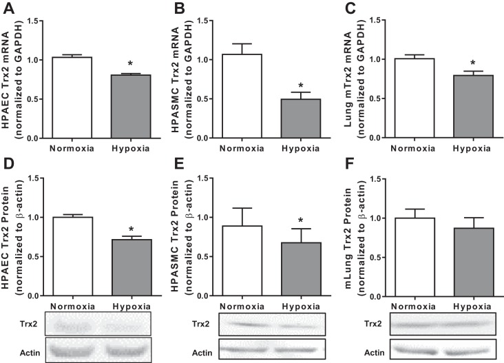
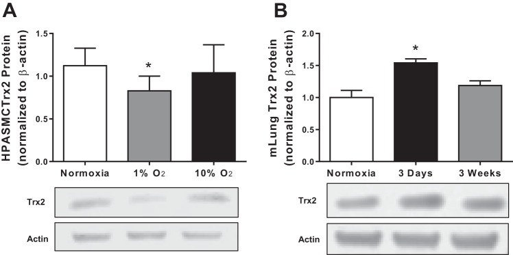
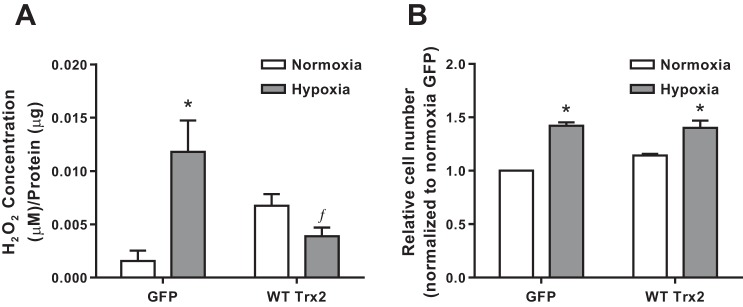
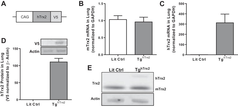
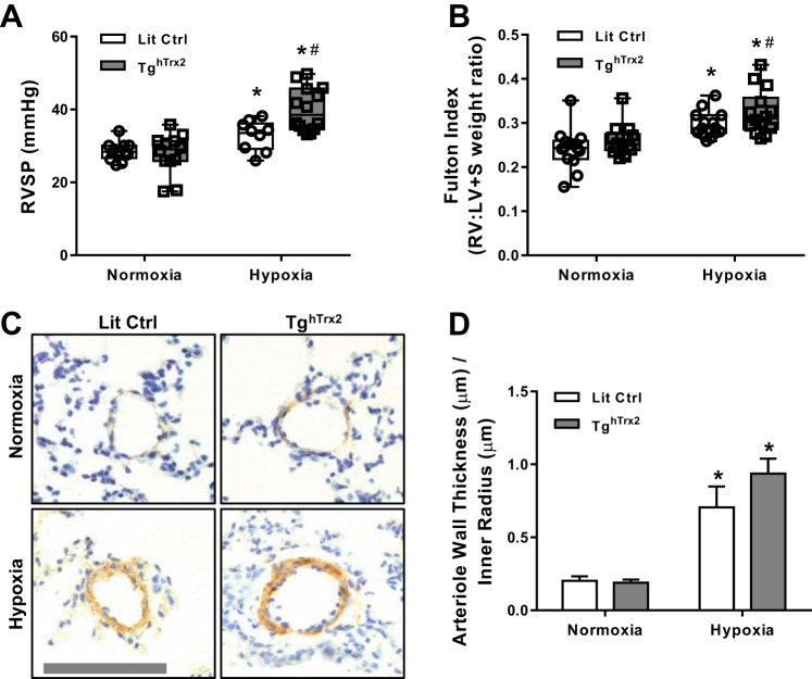
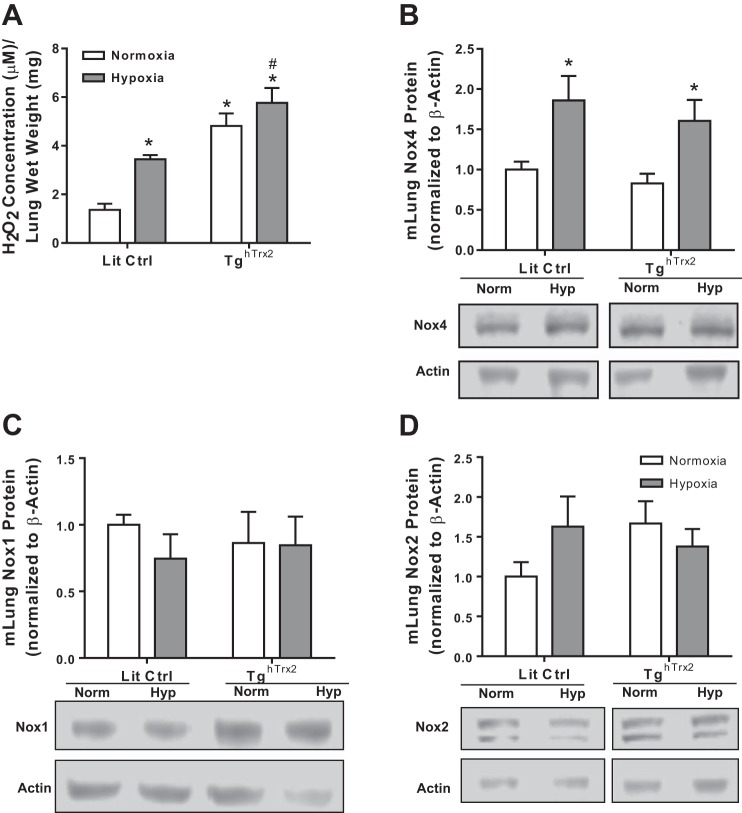
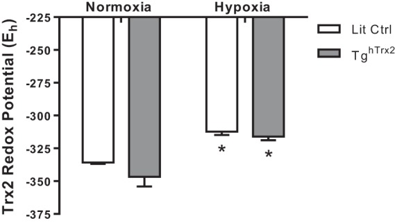
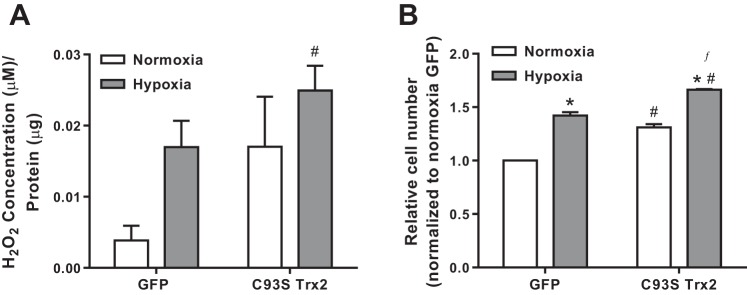
Pulmonary hypertension (PH) is characterized by increased pulmonary vascular resistance, pulmonary vascular remodeling, and increased pulmonary vascular pressures that often result in right ventricular dysfunction, leading to right heart failure. Evidence suggests that reactive oxygen species (ROS) contribute to PH pathogenesis by altering pulmonary vascular cell proliferation and intracellular signaling pathways. However, the role of mitochondrial antioxidants and oxidant-derived stress signaling in the development of hypoxia-induced PH is largely unknown. Therefore, we examined the role of the major mitochondrial redox regulator thioredoxin 2 (Trx2). Levels of Trx2 mRNA and protein were examined in human pulmonary arterial endothelial cells (HPAECs) and smooth muscle cells (HPASMCs) exposed to hypoxia, a common stimulus for PH, for 72 h. Hypoxia decreased Trx2 mRNA and protein levels. In vitro overexpression of Trx2 reduced hypoxia-induced H2O2 production. The effects of increased Trx2 protein level were examined in transgenic mice expressing human Trx2 (TghTrx2) that were exposed to hypoxia (10% O2) for 3 wk. TghTrx2 mice exposed to hypoxia had exacerbated increases in right ventricular systolic pressures, right ventricular hypertrophy, and increased ROS in the lung tissue. Trx2 overexpression did not attenuate hypoxia-induced increases in Trx2 oxidation or Nox4 expression. Expression of a dominant negative C93S Trx2 mutant that mimics Trx2 oxidation exacerbated hypoxia-induced increases in HPASMC H2O2 levels and cell proliferation. In conclusion, Trx2 overexpression failed to attenuate hypoxia-induced HPASMC proliferation in vitro or hypoxia-induced PH in vivo. These findings indicate that strategies to enhance Trx2 expression are unlikely to exert therapeutic effects in PH pathogenesis.

Keywords: human pulmonary artery smooth muscle cell; hydrogen peroxide; pulmonary hypertension; reactive oxygen species.


Figures

Fig. 1.
Fig. 1.
Trx2 expression is decreased in in vitro and in vivo models of hypoxia-induced pulmonary hypertension (PH). Trx2 mRNA normalized to GAPDH (fold change) in HPAECs (n = 3; A) and HPASMCs (n = 6; B) exposed to 72 h of hypoxia and mouse lung (n = 6; C) mRNA after 3 wk of hypoxia (10% O2) or normoxia (21% O2) exposure. Trx2 protein normalized to β-actin (fold change) in HPAECs (n = 5; D), HPASMCs (n = 5; E), and mouse lung (n = 5; F) protein. Trx2, thioredoxin 2; HPAECs, human pulmonary artery endothelial cells; HPASMCs, human pulmonary artery smooth muscle cells; mlung, mouse lung. Error bars = SE. *P < 0.05.
Fig. 2:
Fig. 2:
Trx2 expression is sensitive to fluctuations in oxygen levels. Trx2 protein normalized to β-actin (fold change) in HPASMCs (n = 6; A) exposed to either 1 or 10% O2 and mouse lung (n = 5; B) exposed to either 3 days or 3 wk of hypoxia (10% O2). Error bars = SE. *P < 0.05, compared with Normoxia.
Fig. 3:
Fig. 3:
WT Trx2 overexpression in HPASMCs prevents hypoxia-induced ROS production but not proliferation. A; extracellular H2O2 levels in HPASMCs infected with either GFP control or wild-type Trx2 (WT Trx2) (n ≥ 4). B: cellular proliferation measured in HPASMCs infected with either GFP control or WT Trx2 (n = 3). ROS, reactive oxygen species; WT, wild type; GFP, green fluorescent protein. Error bars = SE. *P < 0.05, compared with Normoxia of same group; ƒP < 0.05, compared with Control Hypoxia).
Fig. 4:
Fig. 4:
Human Trx2 overexpression in the TghTrx2 mouse model. A: schematic depicting Trx2 overexpression construct used to generate TghTrx2 mouse model. B: mouse Trx2 mRNA levels in Lit Ctrl or TghTrx2 (n = 6). Human Trx2 mRNA (n = 3; C) and protein (detected by V5 epitope) (n = 3; D) levels in Lit Ctrl and TghTrx2 animals. E: representative images of mouse (bottom) and human (top) Trx2 protein levels in Lit Ctrl and TghTrx2 animals. Lit Ctrl, littermate control. Error bars = SE.
Fig. 5:
Fig. 5:
Overexpression of Trx2 does not ameliorate physiological markers of hypoxia-induced PH. RVSP (n ≥ 12; A), Fulton Index (n = 14; B), pulmonary vessel muscularization measured by smooth muscle actin staining of vessels smaller than 100 µm in Lit Ctrl and TghTrx2 mice exposed to Hypoxia (10% O2) or Normoxia (21% O2) for 3 wk (n = 3; C and D). RVSP, right ventricular systolic pressures; Fulton index, right ventricle hypertrophy. Error bars = SE. *P < 0.05, compared with Normoxia groups; #P < 0.05, compared with Lit Ctrl Hypoxia.
Fig. 6:
Fig. 6:
Trx2 Overexpression does not prevent hypoxia-induced increase in Nox4. H2O2 production (n ≥ 5; A), Nox 4 protein levels (n = 8; B), Nox 1 (n = 8; C), or Nox2 (n = 8; D) protein levels in Lit Ctrl and TghTrx2 mice exposed to Hypoxia (10% O2) or Normoxia (21% O2) for 3 wk. Nox4, NAD(P)H oxidase 4; Nox1, NAD(P)H oxidase 1; Nox2, NAD(P)H oxidase 2. Error bars = SE. *P < 0.05, compared with Normoxia groups; #P < 0.05, compared with Lit Ctrl Hypoxia.
Fig. 7:
Fig. 7:
Hypoxia decreases Trx2 redox potential in vivo. Trx2 Redox state was calculated using the Nernst equation in conjunction with densitometry analysis of native gels. Error bars = SE. *P < 0.05, compared with Normoxia of same group.
Fig. 8:
Fig. 8:
Trx2 activity is important for preventing hypoxia-induced ROS production and proliferation in HPASMC. A: extracellular H2O2 levels in HPASMC expressing either GFP control or C93S Trx2 (n ≥ 7). B: cellular proliferation measured in HPASMC expressing either GFP control or C93S Trx2 (n = 3). Error bars = SE. *P < 0.05, compared with normoxia of same group; #P < 0.05, compared with GFP Normoxia; ƒP < 0.05, compared with GFP Hypoxia.

References

    1. Adesina SE, Kang BY, Bijli KM, Ma J, Cheng J, Murphy TC, Michael Hart C, Sutliff RL. Targeting mitochondrial reactive oxygen species to modulate hypoxia-induced pulmonary hypertension. Free Radic Biol Med 87: 36–47, 2015. doi:10.1016/j.freeradbiomed.2015.05.042. - DOI - PMC - PubMed
    1. Bijli KM, Kleinhenz JM, Murphy TC, Kang BY, Adesina SE, Sutliff RL, Hart CM. Peroxisome proliferator-activated receptor gamma depletion stimulates Nox4 expression and human pulmonary artery smooth muscle cell proliferation. Free Radic Biol Med 80: 111–120, 2015. doi:10.1016/j.freeradbiomed.2014.12.019. - DOI - PMC - PubMed
    1. Bowers R, Cool C, Murphy RC, Tuder RM, Hopken MW, Flores SC, Voelkel NF. Oxidative stress in severe pulmonary hypertension. Am J Respir Crit Care Med 169: 764–769, 2004. doi:10.1164/rccm.200301-147OC. - DOI - PubMed
    1. Brennan LA, Steinhorn RH, Wedgwood S, Mata-Greenwood E, Roark EA, Russell JA, Black SM. Increased superoxide generation is associated with pulmonary hypertension in fetal lambs: a role for NADPH oxidase. Circ Res 92: 683–691, 2003. doi:10.1161/01.RES.0000063424.28903.BB. - DOI - PubMed
    1. Chen B, Nelin VE, Locy ML, Jin Y, Tipple TE. Thioredoxin-1 mediates hypoxia-induced pulmonary artery smooth muscle cell proliferation. Am J Physiol Lung Cell Mol Physiol 305: L389–L395, 2013. doi:10.1152/ajplung.00432.2012. - DOI - PMC - PubMed

Publication types

MeSH terms

LinkOut - more resources