. 2017 Apr;16(4):552-566.
doi: 10.1074/mcp.M116.063321. Epub 2017 Jan 27.

Melt With This Kiss: Paralyzing and Liquefying Venom of The Assassin Bug Pristhesancus plagipennis (Hemiptera: Reduviidae)

Affiliations

Melt With This Kiss: Paralyzing and Liquefying Venom of The Assassin Bug Pristhesancus plagipennis (Hemiptera: Reduviidae)

Andrew A Walker et al. Mol Cell Proteomics. 2017 Apr.

Abstract

Assassin bugs (Hemiptera: Heteroptera: Reduviidae) are venomous insects, most of which prey on invertebrates. Assassin bug venom has features in common with venoms from other animals, such as paralyzing and lethal activity when injected, and a molecular composition that includes disulfide-rich peptide neurotoxins. Uniquely, this venom also has strong liquefying activity that has been hypothesized to facilitate feeding through the narrow channel of the proboscis-a structure inherited from sap- and phloem-feeding phytophagous hemipterans and adapted during the evolution of Heteroptera into a fang and feeding structure. However, further understanding of the function of assassin bug venom is impeded by the lack of proteomic studies detailing its molecular composition.By using a combined transcriptomic/proteomic approach, we show that the venom proteome of the harpactorine assassin bug Pristhesancus plagipennis includes a complex suite of >100 proteins comprising disulfide-rich peptides, CUB domain proteins, cystatins, putative cytolytic toxins, triabin-like protein, odorant-binding protein, S1 proteases, catabolic enzymes, putative nutrient-binding proteins, plus eight families of proteins without homology to characterized proteins. S1 proteases, CUB domain proteins, putative cytolytic toxins, and other novel proteins in the 10-16-kDa mass range, were the most abundant venom components. Thus, in addition to putative neurotoxins, assassin bug venom includes a high proportion of enzymatic and cytolytic venom components likely to be well suited to tissue liquefaction. Our results also provide insight into the trophic switch to blood-feeding by the kissing bugs (Reduviidae: Triatominae). Although some protein families such as triabins occur in the venoms of both predaceous and blood-feeding reduviids, the composition of venoms produced by these two groups is revealed to differ markedly. These results provide insights into the venom evolution in the insect suborder Heteroptera.


Figures

Fig. 1.
Fig. 1.
A, assassin bug P. plagipennis envenomating feeder cricket (A. domestica). B, extraction of venom from P. plagipennis using electrostimulation.
Fig. 2.
Fig. 2.
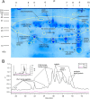
Proteins detected by LC-MS/MS of 2D SDS-PAGE spots and HPLC fractions showing abundant proteases, CUB-domain proteins, and heteropteran venom family 1 proteins. A, 2D SDS-polyacrylamide gel of crude venom, showing protein families identified by LC-MS/MS of gel spots. B, HPLC trace of venom fractionation, showing protein families identified by LC-MS/MS of collected fractions.
Fig. 3.
Fig. 3.
Proportion of sequences belonging to each major protein class in the venom of P. plagipennis.
Fig. 4.
Fig. 4.
Evolution of Ptu1-like peptides isolated from P. plagipennis venom. Signal sequences predicted by SignalP4.1 are shown in lowercase; lines above the text show the disulfide connectivity for Ptu1. Sequence labels in blue indicate peptides detected in venom using proteomics. POI, phenoloxidase inhibitor; AMP, antimicrobial peptide. Node labels indicate posterior probabilities. Comparison sequences are remipede (S. tulumensis) agatoxin-like peptide (66), assassin bug venom peptides Ado1 (P58608.1), Iob1 (P58609.1), and Ptu1 (P58606.1); bee (Apis mellifera) OCLP1 (H9KQJ7.1); ant (Trachymyrmex cornetzi) predicted protein (KYN16117.1); ant (Wasmannia auropunctata) conotoxin-like peptide (XP_011706926.1); fly (Bradysia hygida) salivary putative AMP1 (ABA26621.1); and Hemiptera (Bemisia tabaci) putative AMP knottin peptide Btk-1 (ABC40569.1).
Fig. 5.
Fig. 5.
Evolution of CUB domain proteins. Sequence names in blue indicate P. plagipennis sequences determined in this study. A, alignment of CUB domain venom proteins from P. plagipennis and the anthocorid O. laevigatus (52) with the N-terminal portion (including CUB domain) of P. plagipennis venom S1 protease 8. B, phylogeny according to Bayesian inference showing support for monophyly of cimicomorphan venom CUB domain proteins (highlighted yellow). Anthocorid CUB domain protein, GBG01000009.1; crustacean CUB-S1 protease, AAK48894.1; beetle CUB-S1 protease, ENN74674.1; mosquito CUB-S1 protease, XP_001657965.1.
Fig. 6.
Fig. 6.
Conserved sequence features in redulysin proteins. A, amino acid sequence alignment of redulysins with the cytolytic domain of trialysin (AAL82381.1). The consensus sequence of the redulysin proteins is shown below. Lys residues are shown in bold black, positively charged residues in red, and hydrophobic residues in blue. Residues predicted by PSIPRED to form α-helices are highlighted in gray. B, helical wheel diagram of the predicted helical portion of the consensus of redulysin sequences showing clustering of Lys residues and hydrophobic residues on opposite sides of the helix.
Fig. 7.
Fig. 7.
Radiation of triabin/lipocalin superfamily in Reduviidae and allied insects calculated by Bayesian inference. Branches of Pristhesancus, Triatoma, and Rhodnius proteins are colored orange, blue, and pink, respectively; node labels indicate posterior probabilities. Accessions of sequences used in this analysis are as follows: putative BABP (Triatoma), D1MX91; putative nitrophorin (Triatoma), A0A023F6B8; Triafestin, AB292809.1; RPAI, JA76747.1; Triplatin, BAE96121.1; Infestilin, AAZ38958.1; Pallidipin, AAA30329.1; Dimiconin, BAI50848.1; Triabin, CAA56540.1; Procalin, AEM97970.1; Triatin, AAZ38956.1; ApoD-like (Halyomorpha), XP_014291570.1; Isoallergen 1 (Blatella), C3RWZ4; ApoD-like (Cimex), XP_014256007.1; Uncharacterized (Cimex), XP_014250080.1; Lazarillo-like (Cimex), XP_014249890; Lipocalin (Homo), CAA47889.1.

References

    1. Fry B. G., Roelants K., Champagne D. E., Scheib H., Tyndall J. D., King G. F., Nevalainen T. J., Norman J. A., Lewis R. J., Norton R. S., Renjifo C., and de la Vega R. C. (2009) The toxicogenomic multiverse: convergent recruitment of proteins into animal venoms. Annu. Rev. Genomics Hum. Genet. 10, 483–511 - PubMed
    1. Olivera B. M., Walker C., Cartier G. E., Hooper D., Santos A. D., Schoenfeld R., Shetty R., Watkins M., Bandyopadhyay P., and Hillyard D. R. (1999) Speciation of cone snails and interspecific hyperdivergence of their venom peptides. Ann. N.Y. Acad. Sci. 870, 223–237 - PubMed
    1. Sollod B. L., Wilson D., Zhaxybayeva O., Gogarten J. P., Drinkwater R., and King G. F. (2005) Were arachnids the first to use combinatorial peptide libraries? Peptides 26, 131–139 - PubMed
    1. Sunagar K., Jackson T. N., Undheim E. A., Ali S. A., Antunes A., and Fry B. G. (2013) Three-Fingered RAVERs: rapid accumulation of variations in exposed residues of snake venom toxins. Toxins 5, 2172–2208 - PMC - PubMed
    1. Sunagar K., and Moran Y. (2015) The rise and fall of an evolutionary innovation: contrasting strategies of venom evolution in ancient and young animals. PLoS Genet. 11, e1005596. - PMC - PubMed

Publication types

Substances

LinkOut - more resources