. 2017 Jun;28(6):1769-1782.
doi: 10.1681/ASN.2016080826. Epub 2017 Jan 27.

Connective Tissue Growth Factor Domain 4 Amplifies Fibrotic Kidney Disease through Activation of LDL Receptor-Related Protein 6

Affiliations

Connective Tissue Growth Factor Domain 4 Amplifies Fibrotic Kidney Disease through Activation of LDL Receptor-Related Protein 6

Bryce G Johnson et al. J Am Soc Nephrol. 2017 Jun.

Abstract

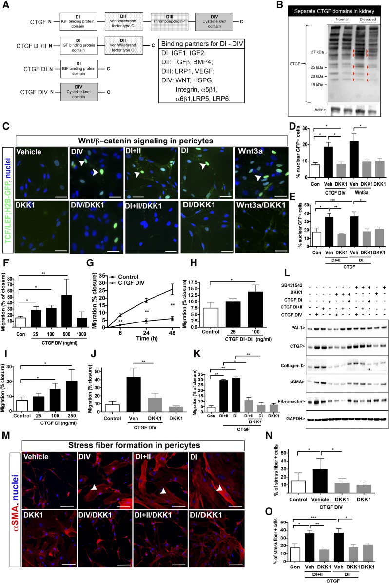
Connective tissue growth factor (CTGF), a matrix-associated protein with four distinct cytokine binding domains, has roles in vasculogenesis, wound healing responses, and fibrogenesis and is upregulated in fibroblasts and myofibroblasts in disease. Here, we investigated the role of CTGF in fibrogenic cells. In mice, tissue-specific inducible overexpression of CTGF by kidney pericytes and fibroblasts had no bearing on nephrogenesis or kidney homeostasis but exacerbated inflammation and fibrosis after ureteral obstruction. These effects required the WNT receptor LDL receptor-related protein 6 (LRP6). Additionally, pericytes isolated from these mice became hypermigratory and hyperproliferative on overexpression of CTGF. CTGF is cleaved in vivo into distinct domains. Treatment with recombinant domain 1, 1+2 (N terminus), or 4 (C terminus) independently activated myofibroblast differentiation and wound healing responses in cultured pericytes, but domain 4 showed the broadest profibrotic activity. Domain 4 exhibited low-affinity binding to LRP6 in in vitro binding assays, and inhibition of LRP6 or critical signaling cascades downstream of LRP6, including JNK and WNT/β-catenin, inhibited the biologic activity of domain 4. Administration of blocking antibodies specifically against CTGF domain 4 or recombinant Dickkopf-related protein-1, an endogenous inhibitor of LRP6, effectively inhibited inflammation and fibrosis associated with ureteral obstruction in vivo Therefore, domain 4 of CTGF and the WNT signaling pathway are important new targets in fibrosis.

Keywords: Connective Tissue Growth Factor; DKK1; LRP6; fibrosis; inflammation; pericytes.


Figures

Figure 1.
Figure 1.
Conditional CTGF overexpression in kidney stromal cells exacerbates fibrotic and inflammatory responses and requires LRP6. (A) Gene map of Foxd1Cre and R26-CIG transgenes. (B) Images of adult kidney showing Foxd1 nephrogenic progenitor–derived resident pericytes (upper panel; arrows) in the medulla and mesangial cells (arrowheads; lower panel) in the glomerulus (g) overexpressing CTGF detected by nGFP and coexpressing PDGFRβ. Podocytes also activate Foxd1 during maturation and therefore, overexpress CTGF identified with nGFP (arrows; lower panel). (C) Histologic staining of control and CTGF overexpressing tissues at baseline revealing no significant difference in periodic acid–Schiff (PAS) and picrosirius red stains. (D) Graph showing morphometric results of immunofluorescence labeling of healthy adult kidneys to identify pericytes (PDGFRβ), activated fibroblasts/myofibroblasts (αSMA), and endothelial cells (CD31). (E) Transcriptional analysis of healthy adult kidney tissue for genes encoding extracellular matrix components, growth factors, and cytokines normalized to control samples. (F) Schema showing surgical induction of kidney disease and timeline. (G) Immunofluorescence labeling to identify pericytes (PDGFRβ) and macrophages (F4/80) and histologic stain for fibrillar collagen (picrosirius red) 7 days after UUO. (H) Graphs showing morphometric analysis for extracellular matrix components, macrophages, interstitial fibroblasts, and fibrillar collagen deposition. (I and J) Transcriptional analysis of kidney tissue of genes encoding extracellular matrix components, growth factors, and cytokines normalized to control samples at baseline and disease conditions. (K) Transcript levels for WNT downstream target genes in kidney tissue. (L and M) Levels of extracellular matrix and inflammatory gene transcription in Foxd1Cre;R26-CIG kidney in response to Lrp6 antisense gapmer oligonucleotide (ASO) treatment. Data are represented as mean±SEM; n=4–7 per group. Scale bars, 50 µm. *P<0.05; **P<0.01; ***P<0.001; ****P<0.001.
Figure 2.
Figure 2.
Distinct domains of CTGF activate WNT/β-catenin in pericytes, which is inhibited by DKK1. (A) Schema showing domains of CTGF and potential binding partners. (B) Western blot detecting CTGF and its domains in total kidney (normal kidney/diseased kidney). Note that, in diseased kidneys, the putative cleaved CTGF fragment bands are shown with arrowheads. (C) Images and (D and E) graphs showing that CTGF domains activate WNT signaling in mouse pericytes as detected by nGFP 16 hours after activation in TCF/LEF:H2B-GFP transgenic pericytes. Recombinant DKK1 inhibits distinct CTGF domain–induced canonical WNT responses in mouse pericytes. (F and G) Graphs showing that CTGF DIV induces pericyte migration in a time- and dose-dependent manner. (H and I) CTGF DI+II and DI alone stimulate migration with dose dependency. (J) CTGF DIV–induced migration is blocked by recombinant DKK1 (K) CTGF DI+II– or DI–induced migration is blocked by recombinant DKK1. (L) Western blots showing CTGF DIV– and CTGF DI+II–induced matrix proteins, αSMA, and CTGF. DKK1 inhibits all responses to DIV and most responses to DI+II. SB431542 has weak effects on matrix responses only. (M–O) Images and graphs showing the effect of CTGF domains on stress fiber formation (arrowheads). DKK1 blocks responses to each domain. Data are mean±SEM; n=6 per group. All blots are representative of three experiments. Scale bars, 25 μm. *P<0.05; **P<0.01; ***P<0.001.
Figure 3.
Figure 3.
CTGF domain 4 directly activates the WNT coreceptor LRP6 and stimulates migration predominantly by JNK phosphorylation in pericytes. (A) Western blots detecting pLRP6 or CTGF among mouse pericyte proteins immunoprecipitated by anti-pLRP6 antibodies or control antibodies after stimulation with CTGF DIV with or without DKK1. (B) Western blots of phosphorylated forms of P42/P44 MAPK, JNK, P38 MAPK, and LRP6 in pericytes activated by CTGF DIV or CTGF DIV and DKK1. (C) CTGF DIV–mediated migration is blocked by the JNK inhibitor (SP600125) and WNT export inhibitor (IWP2) but is not blocked by the β-catenin inhibitor (XAV939), TGFβRI inhibitor (SB431542), or P42/44 inhibitor (U0126) in quiescent pericytes at 24 hours. (D) Western blots of phosphorylated forms of P42/P44, JNK, P38, and LRP6 in pericytes activated by CTGF DIV, DI, or DI+II. (E and F) Graphs showing that migration in response to both CTGF DI+II and CTGF DI alone is selectively blocked by the JNK inhibitor (SP600125) and WNT export inhibitor (IWP2) in pericytes after 24 hours. (G–I) Western blots and graphs showing the effect of siRNA against LRP6 or β-catenin or scrambled sequence on expression of LRP6 and β-catenin and the subsequence effect on migration triggered by CTGF domains. (J, K) Blots showing the effect of signaling responses to CTGF domains after silencing of LRP6 or mock silencing in basal/unstimulated conditions (J) or in conditions stimulated by different CTGF domains (K). (L) The effect of blocking antibodies against CTGF DIV on migration induced by CTGF domains. (M) The effect of antibodies against CTGF DIV on DIV-induced PAI-1 expression. Data are expressed as mean±SEM; n=6 per group. Blots are representative of three independent experiments. *P<0.05; **P<0.01; ***P<0.001.
Figure 4.
Figure 4.
Domain 4 of CTGF causes migration and stress fiber formation and dominantly induces profibrotic responses in human kidney pericytes, which are blocked by DKK1. (A) Flow cytometric plot and immunofluorescence images indicating selection of NG2+ PDGFRβ+ cells from human fetal kidney results in a population of cells that are PDGFRβ+, transiently NG2+, and weakly αSMA+. (B) Migratory responses to CTGF domains at 24 hours in human are all inhibited by DKK1. (C and D) Stress fibers form in response to CTGF domains and are all blocked by DKK1. (E) Quantitative PCR indicates that CTGF domain 4 alone upregulates transcripts associated with fibrogenesis 48 hours after stimulation. (F) Western blots showing COL1, PAI-1, and CTGF production in response to a number of stimuli at 24 hours. Note that DIV protein markedly increases these proteins, whereas DI+II is less effective, and DI alone produces no response. DKK1 blocks these responses, and SB431542 also inhibits PAI-1 and CTGF responses. (G and H) Blots showing signaling responses of the different CTGF domains in human pericytes. Data are mean±SEM; n=6 per group. Blots are representative of three independent experiments. *P<0.05; **P<0.01; ***P<0.001.
Figure 5.
Figure 5.
DKK1 or anti-CTGF domain 4 antibodies inhibit fibrogenic signaling in models of kidney disease. (A) Schema showing the timeline for UUO-mediated disease induction and the delivery of rDKK1 or vehicle. (B) Western blot showing total DKK1 levels in kidney tissue after disease induction and delivery of vehicle or rDKK1. (C) Graph showing the effect of rDKK1 on expression of WNT/β-catenin responses in kidney after UUO. (D) Representative images and (E) morphometric quantification of myofibroblasts and interstitial fibrosis 7 days after UUO. (F) Western blots showing CTGF-responsive protein levels in the kidney in response to rDKK1. (G) Quantitative PCR for levels for transcripts of matrix proteins, fibrogenic factors, and inflammatory response factors. (H) Quantification of F4/80+ macrophages after 7 days of UUO. (I) Schema showing the timeline for UUO-mediated disease induction and the delivery of anti-CTGF domain 4 antibodies or vehicle. (J) Effect of anti-CTGF DIV on WNT/β-catenin responses in kidney after UUO. (K) Representative images and (L) quantification of laminin accumulation and interstitial fibrosis. (M) Graphs showing morphometric quantification of protein markers in kidney tissue sections in response to disease and anti-CTGF DIV antibodies. (N) Western blots showing CTGF-responsive protein levels in the kidney in response to anti-CTGF DIV. (O) Quantitative PCR for transcript levels of matrix proteins and inflammatory factors in response to (L) anti-CTGF DIV. Blots are representative of two or more independent experiments; n=6 per group. Scale bar, 50 μm. *P<0.05; **P<0.01; ***P<0.001; ****P<0.0001.

References

    1. Lupher ML Jr., Gallatin WM: Regulation of fibrosis by the immune system. Adv Immunol 89: 245–288, 2006 - PubMed
    1. Duffield JS: Cellular and molecular mechanisms in kidney fibrosis. J Clin Invest 124: 2299–2306, 2014 - PMC - PubMed
    1. Humphreys BD, Lin SL, Kobayashi A, Hudson TE, Nowlin BT, Bonventre JV, Valerius MT, McMahon AP, Duffield JS: Fate tracing reveals the pericyte and not epithelial origin of myofibroblasts in kidney fibrosis. Am J Pathol 176: 85–97, 2010 - PMC - PubMed
    1. Kramann R, Schneider RK, DiRocco DP, Machado F, Fleig S, Bondzie PA, Henderson JM, Ebert BL, Humphreys BD: Perivascular Gli1+ progenitors are key contributors to injury-induced organ fibrosis. Cell Stem Cell 16: 51–66, 2015 - PMC - PubMed
    1. Schrimpf C, Xin C, Campanholle G, Gill SE, Stallcup W, Lin SL, Davis GE, Gharib SA, Humphreys BD, Duffield JS: Pericyte TIMP3 and ADAMTS1 modulate vascular stability after kidney injury. J Am Soc Nephrol 23: 868–883, 2012 - PMC - PubMed

MeSH terms

Substances