. 2017 Jul;134(1):65-78.
doi: 10.1007/s00401-017-1679-9. Epub 2017 Jan 27.

Expansion of the classification of FTLD-TDP: distinct pathology associated with rapidly progressive frontotemporal degeneration

Affiliations

Expansion of the classification of FTLD-TDP: distinct pathology associated with rapidly progressive frontotemporal degeneration

Edward B Lee et al. Acta Neuropathol. 2017 Jul.

Abstract

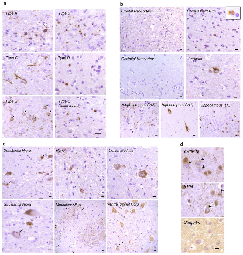
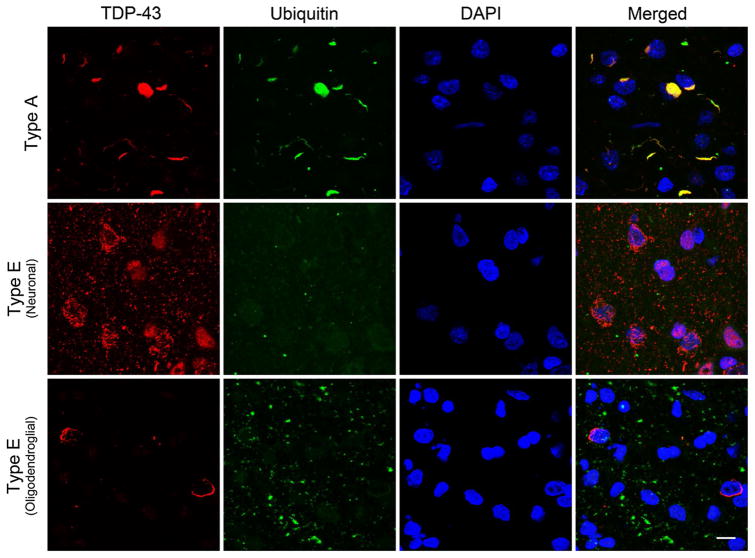
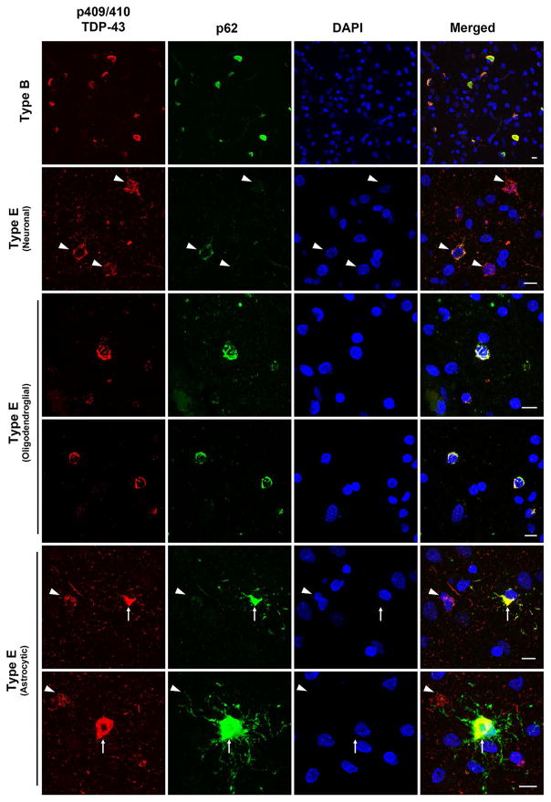
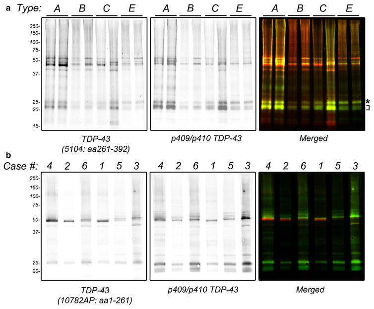
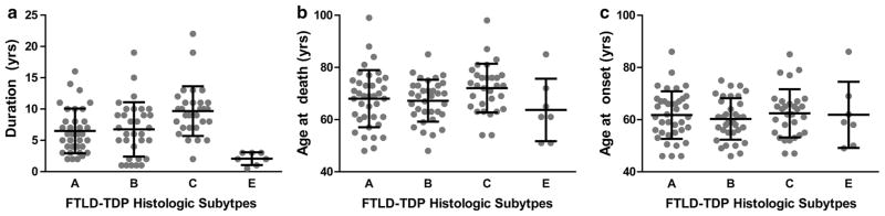
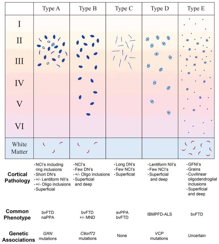
Frontotemporal lobar degeneration with TDP-43 inclusions (FTLD-TDP) can typically be categorized into one of four distinct histopathologic patterns of TDP-43 pathology, types A to D. The strength of this histopathologic classification lies in the association between FTLD-TDP subtypes and various clinical and genetic features of disease. Seven cases of FTLD-TDP were identified here which were difficult to classify based on existing pathologic criteria. Distinct features common to these cases included TDP-43 aggregates over a wide neuroanatomic distribution comprised of granulofilamentous neuronal inclusions, abundant grains, and oligodendroglial inclusions. TDP-43 aggregates were phosphorylated and associated with loss of normal nuclear TDP-43 protein (nuclear clearance) but were negative for ubiquitin. Biochemical analysis confirmed the presence of insoluble and phosphorylated TDP-43 and also revealed a distinct pattern of TDP-43 C-terminal fragments relative to other FTLD-TDP subtypes. Finally, these cases were uniformly associated with a very rapid clinical course culminating in death within ~3 years of disease onset. We suggest that these cases may represent a unique clinicopathologic subtype of FTLD-TDP which we provisionally call "type E." The immature appearance of TDP-43 aggregates, widespread distribution, uniform biochemical profile and rapid clinical course highlights the clinical and pathologic variability within FTLD-TDP, and raises the possibility that type E neuropathology is the sequelae of a particularly virulent strain of TDP-43 proteinopathy.


Figures

Fig. 1
Fig. 1
Histopathologic characteristics of FTLD-TDP type E. a Representative images of p409/p410 immunostained neocortical sections (layer II) from type A, B, C, D and E are shown (scale bar 20 μm). Type A shows dense neuronal cytoplasmic inclusions including ring inclusions, and neurites affecting superficial neocortical layers. Type B shows compact neuronal cytoplasmic inclusions with few neurites and involves superficial and deep neocortical layers. Type C shows long dystrophic neurites. Type D is typified by numerous intranuclear inclusions. Type E exhibits GFNI’s and grains in superficial and deep neocortical layers. Curvilinear oligodendroglial inclusions are also present (white matter). b Type E pathology is seen throughout most of the cerebrum including frontal neocortex, major white matter tracts such as the corpus callosum (inset shows high power image of curvilinear oligodendroglial inclusion), deep grey structures such as the striatum, and limbic regions such as the hippocampus (DG = dentate gyrus). Also shown is the occipital neocortex which is relatively spared (scale bars 10 μm). c Type E pathology is seen throughout the brainstem and cervical spinal cord. Shown are representative images from the substantia nigra, basis pontis (pons), medullary olive, dorsal medulla and ventral spinal cord. More compact or dense inclusions highlighted with arrows. Oligodendroglial inclusions highlighted with asterisks. Scale bars 10 μm. d Representative images of type E cases stained with anti-TDP-43 antibodies 6H6E12 and 5104, and the anti-ubiquitin antibody MAB1510 are shown (scale bar 10 μm). TDP-43 antibodies reveal both GFNI’s and grains with nuclear clearance of normal nuclear TDP-43 in affected neurons (open arrowheads). Ubiquitin stain was negative for discernible inclusions
Fig. 2
Fig. 2
TDP-43 positive but ubiquitin-negative inclusions in FTLD-TDP type E. Representative confocal stack images are shown of double immunofluorescence stained cortical sections stained using anti-TDP-43 (C1039, rabbit polyclonal, red) and anti-ubiquitin (MAB1510, mouse monoclonal, green) with a DAPI nuclear counterstain (blue). A typical type A case exhibits inclusions positive for both ubiquitin and TDP-43 positive inclusions (top panels). In contrast, neuronal GFNI’s and grains from a type E case are TDP-43 positive but ubiquitin negative (middle panel). Similarly, curvilinear oligodendroglial inclusions are also TDP-43 positive but ubiquitin negative (bottom panel). Scale bar 10 μm
Fig. 3
Fig. 3
TDP-43 positive and variably p62-positive inclusions in FTLD-TDP type E. Representative confocal stack images are shown of double immunofluorescence stained cortical sections stained using anti-p409/410 TDP-43 (TIP-PTD-P01, rabbit polyclonal, red) and anti-p62 (2C11, mouse monoclonal, green) with a DAPI nuclear counterstain (blue). A typical type B case exhibits inclusions positive for both p62 and TDP-43 positive inclusions (top panels). In contrast, neuronal GFNI’s and grains from a type E case are TDP-43 positive but variably positive for p62 with a small minority of GFNI’s exhibiting moderate p62 staining (upper middle panels). Curvilinear oligodendroglial inclusions (middle panels) and astrocytic inclusion (bottom panels) were strongly positive for phospho-TDP-43 and p62. GNFI’s are highlighted with arrowheads. Astrocytic inclusions are highlighted with arrows. Scale bars 10 μm
Fig. 4
Fig. 4
Biochemical analysis of FTLD-TDP type E. Frozen frontal cortex from unfixed frozen human brain was subjected to sequential biochemical extraction using a series of buffers of increasing strength. Sarkosyl-insoluble fractions were electrophoresed on NuPage Bis–Tris gels and immunoblotted with primary antibodies as indicated and detected using fluorescently-labeled secondary antibodies followed by dual-color imaging. a Representative immunoblot of type A, B, C and E cases using 5104 (anti-TDP-43 antibody that recognizes amino acids 261–392, left panel and red in right panel) and TIP-PTD-P01 (anti-p409/p410-TDP-43 antibody that recognizes phosphorylated S409 and S410, middle panel and green in right panel). Asterisk highlights upper C-terminal fragment band. Bracket highlights smaller C-terminal fragments. Sufficient numbers of type D cases were not available for analysis. b Immunoblotting of six type E cases (labeled according to Table 1) with 10782-AP (anti-TDP-43 antibody raised against amino acids 1-261, left panel and red in right panel) and 1D3 (anti-p409/p410-TDP-43, middle panel and green in right panel)
Fig. 5
Fig. 5
FTLD-TDP Subtypes and disease duration, age at death and age at onset. a Disease duration, b age at death and c age at onset are shown for FTLD-TDP type A, B, C and E cases. Data presented for each individual together with group mean (lines) and standard deviation (error bars). Sufficient numbers of type D cases were not available for analysis
Fig. 6
Fig. 6
Proposed expansion of FTLD-TDP classification. An illustration of TDP-43 aggregate morphology that define Types A, B, C, D, and E is shown. Below each FTLD-TDP type is a summary of the pathologic features, clinical phenotypes, and genetic features of each subtype. Type A is characterized by crescentic to oval/ring-like neuronal cytoplasmic inclusions and many short dystrophic neurites involving superficial neocortical layers. Lentiform neuronal intra-nuclear inclusions and oligodendroglial (oligo) inclusions may also be observed. Type B is characterized by neuronal cytoplasmic inclusions affecting superficial and deep neocortical layers with a paucity of dystrophic neurites. Oligodendroglial inclusions may be observed. Type C is characterized by long dystrophic neurites predominantly in superficial layers with a paucity of neuronal cytoplasmic inclusions. Type D is characterized by frequent lentiform neuronal intranuclear inclusions with short dystrophic neurites. Finally, Type E is characterized by granulofilamentous neuronal inclusions and very fine, dot-like neuropil aggregates affecting all neocortical layers in addition to curvilinear oligodendroglial inclusions in the white matter. Modified from Mackenzie et al. [22]

References

    1. Brettschneider J, Arai K, Del Tredici K, Toledo JB, Robinson JL, Lee EB, Kuwabara S, Shibuya K, Irwin DJ, Fang L, et al. TDP-43 pathology and neuronal loss in amyotrophic lateral sclerosis spinal cord. Acta Neuropathol. 2014;128:423–437. doi: 10.1007/s00401-014-1299-6. - DOI - PMC - PubMed
    1. Brettschneider J, Del Tredici K, Irwin DJ, Grossman M, Robinson JL, Toledo JB, Lee EB, Fang L, Van Deerlin VM, Ludolph AC, et al. Sequential distribution of pTDP-43 pathology in behavioral variant frontotemporal dementia (bvFTD) Acta Neuropathol. 2014;127:423–439. doi: 10.1007/s00401-013-1238-y. - DOI - PMC - PubMed
    1. Brettschneider J, Del Tredici K, Lee VM, Trojanowski JQ. Spreading of pathology in neurodegenerative diseases: a focus on human studies. Nat Rev Neurosci. 2015;16:109–120. doi: 10.1038/nrn3887. - DOI - PMC - PubMed
    1. Brettschneider J, Del Tredici K, Toledo JB, Robinson JL, Irwin DJ, Grossman M, Suh E, Van Deerlin VM, Wood EM, Baek Y, et al. Stages of pTDP-43 pathology in amyotrophic lateral sclerosis. Ann Neurol. 2013;74:20–38. doi: 10.1002/ana.23937. - DOI - PMC - PubMed
    1. Cairns NJ, Bigio EH, Mackenzie IR, Neumann M, Lee VM, Hatanpaa KJ, White CL, 3rd, Schneider JA, Grinberg LT, Halliday G, et al. Neuropathologic diagnostic and nosologic criteria for frontotemporal lobar degeneration: consensus of the consortium for frontotemporal lobar degeneration. Acta Neuropathol. 2007;114:5–22. doi: 10.1007/s00401-007-0237-2. - DOI - PMC - PubMed