Review
. 2017 Mar;67(2):283-301.
doi: 10.1007/s12576-017-0521-4. Epub 2017 Jan 27.

Physiological roles of zinc transporters: molecular and genetic importance in zinc homeostasis

Affiliations
Review

Physiological roles of zinc transporters: molecular and genetic importance in zinc homeostasis

Takafumi Hara et al. J Physiol Sci. 2017 Mar.

Abstract

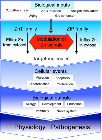
Zinc (Zn) is an essential trace mineral that regulates the expression and activation of biological molecules such as transcription factors, enzymes, adapters, channels, and growth factors, along with their receptors. Zn deficiency or excessive Zn absorption disrupts Zn homeostasis and affects growth, morphogenesis, and immune response, as well as neurosensory and endocrine functions. Zn levels must be adjusted properly to maintain the cellular processes and biological responses necessary for life. Zn transporters regulate Zn levels by controlling Zn influx and efflux between extracellular and intracellular compartments, thus, modulating the Zn concentration and distribution. Although the physiological functions of the Zn transporters remain to be clarified, there is growing evidence that Zn transporters are related to human diseases, and that Zn transporter-mediated Zn ion acts as a signaling factor, called "Zinc signal". Here we describe critical roles of Zn transporters in the body and their contribution at the molecular, biochemical, and genetic levels, and review recently reported disease-related mutations in the Zn transporter genes.

Keywords: Disease; Physiology; Transporter; Zinc; Zinc signaling.


Conflict of interest statement

Author Takafumi Hara declares that he has no conflict of interest. Author Taka-aki Takeda declares that he has no conflict of interest. Author Teruhisa Takagishi declares that he has no conflict of interest. Author Kazuhisa Fukue declares that he has no conflict of interest. Author Taiho Kambe declares that he has no conflict of interest. Author Toshiyuki Fukada declares that he has no conflict of interest.

Figures

Fig. 1
Fig. 1
Zn storage and distribution in the body. Dietary Zn is absorbed from the small intestine and distributed to the organs. Bones and skeletal muscles act as major Zn reservoir tissues
Fig. 2
Fig. 2
Zn storage and distribution in intracellular compartments. The upper diagram shows Zn concentrations in the extracellular region and the cellular compartments (cytosol, mitochondria, ER, and Golgi). The lower diagram shows the direction of Zn transport (black arrows) elicited by ZIP (orange) and ZnT (green) proteins expressed on these cellular compartments
Fig. 3
Fig. 3
The putative structures of ZnT and ZIP transporters. Left side: the putative topology of ZnT transporters. ZnT transporters efflux Zn from the cytosol to the extracellular space or to the lumen of intracellular compartments. ZnT transporters are thought to have six TMDs consisting of two bundles of a compact four-helix (TMDs I, II, IV, and V) and a two-helix pair (TMDs III and VI). They are thought to function as Y-shaped dimers for Zn transport, based on the structural information of E. coli YiiP (shown in top-left panel, PDB 3H90) [–74]. Most ZnT transporters have an indispensable intramembranous Zn-binding site (site A, indicated in magenta circle) consisting of two His (magenta) and two Asp (yellow) residues (HDHD core motif). The position of the His residue (red circle) is speculated to regulate metal substrate specificity. The cytosolic carboxyl-terminal domain (pink square) contains the cytosolic Zn-binding site (site C, indicated in dark green circle), and is thought to consist of two α helixes and three β sheets (αββαβ). The Zn-binding site corresponding to site B in YiiP is omitted because this site is not conserved among ZnT transporters. The cytosolic His-rich loop is indicated in green. The PP motif in the luminal loop in ZnT5 and ZnT7, which is important for TNAP activation [139], is shown in red. Putative Zn chaperon proteins in the cytosol may transfer Zn to the ZnT transporters (see text). Right side: the putative topology of ZIP transporters. This diagram is based on the information available for ZIP4, which is in the LIV-1 subfamily [93, 95]. ZIP transporters mobilize Zn in a direction opposite to that of ZnT transporters. ZIP transporters are thought to have eight TMDs and to function as dimers (not shown). The His residue (magenta) in TMD V is speculated to form part of an intramembranous Zn-binding site, and this position may be involved in specifying the substrate metal. ZIP transporters of the LIV-1 subfamily are characterized by a long extracellular amino-terminal portion containing the helix-rich domain (HRD, orange) and the PAL motif–containing domain (PCD, blue). A potential metalloprotease motif (HEXPHEXGD) is embedded in TM helix V (pale green). Some ZIP transporters have a His cluster (purple) in the cytosolic loop between TMDs III and IV
Fig. 4
Fig. 4
ZnT and ZIP intracellular localizations. The diagram shows the localization of ZnT (green) and ZIP (yellow) proteins, and the direction of Zn transport (black arrows) for each organelle and plasma membrane. In terms of Zn homeostasis, ZnT and ZIP maintain the influx and efflux of Zn ions between the cell and extracellular spaces, or between the cytosol and the organelle compartments, thereby maintaining appropriate Zn concentrations in the cells
Fig. 5
Fig. 5
Summary of Zn transporters in physiology and pathogenesis. Biological inputs such as oxidative stress, antigen stimulation, aging, growth factors, and virus infection trigger various intracellular processes (blue square on upper side). “Modulation of Zn signals” intends the Zn ion, which is transported through individual Zn transporters, modulates various intracellular processes followed by the regulation of molecular status of their target molecules (red arrow area in the middle). Zn signal affects numerous cellular events such as migration, differentiation, proliferation and apoptosis, etc. These cellular events contribute to induce specific biological outputs such as allergy, development, immunity, nerve system and endocrine, etc. (dark blue area on lower side). The impairment of Zn transporter-mediated Zn signal will cause the progression and initiation of various diseases. Please refer to Tables 1 and 2 for reviewing individual biological functions of zinc transporters

References

    1. Andreini C, Bertini I. A bioinformatics view of zinc enzymes. J Inorg Biochem. 2012;111:150–156. doi: 10.1016/j.jinorgbio.2011.11.020. - DOI - PubMed
    1. Andreini C, Bertini I, Rosato A. Metalloproteomes: a bioinformatic approach. Acc Chem Res. 2009;42:1471–1479. doi: 10.1021/ar900015x. - DOI - PubMed
    1. Maret W, Li Y. Coordination dynamics of zinc in proteins. Chem Rev. 2009;109:4682–4707. doi: 10.1021/cr800556u. - DOI - PubMed
    1. Prasad AS. Zinc: an overview. Nutr Burbank Los Angel Cty Calif. 1995;11:93–99. - PubMed
    1. Vallee BL, Auld DS. Cocatalytic zinc motifs in enzyme catalysis. Proc Natl Acad Sci USA. 1993;90:2715–2718. doi: 10.1073/pnas.90.7.2715. - DOI - PMC - PubMed