. 2017 May;12(5):821-828.
doi: 10.1007/s11548-017-1524-6. Epub 2017 Jan 27.

Evaluation of tongue squamous cell carcinoma resection margins using ex-vivo MR

Affiliations

Evaluation of tongue squamous cell carcinoma resection margins using ex-vivo MR

Stefan C A Steens et al. Int J Comput Assist Radiol Surg. 2017 May.

Abstract

Purpose: Purpose of this feasibility study was (1) to evaluate whether application of ex-vivo 7T MR of the resected tongue specimen containing squamous cell carcinoma may provide information on the resection margin status and (2) to evaluate the research and developmental issues that have to be solved for this technique to have the beneficial impact on clinical outcome that we expect: better oncologic and functional outcomes, better quality of life, and lower costs.

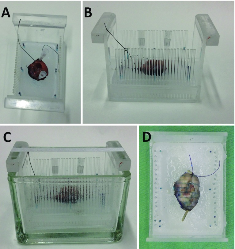
Methods: We performed a non-blinded validation of ex-vivo 7T MR to detect the tongue squamous cell carcinoma and resection margin in 10 fresh tongue specimens using histopathology as gold standard.

Results: In six of seven specimens with a histopathologically determined invasion depth of the tumor of [Formula: see text] mm, the tumor could be recognized on MR, with a resection margin within a 2 mm range as compared to histopathology. In three specimens with an invasion depth of [Formula: see text] mm, the tumor was not visible on MR. Technical limitations mainly included scan time, image resolution, and the fact that we used a less available small-bore 7T MR machine.

Conclusion: Ex-vivo 7T probably will have a low negative predictive value but a high positive predictive value, meaning that in tumors thicker than a few millimeters we expect to be able to predict whether the resection margin is too small. A randomized controlled trial needs to be performed to show our hypothesis: better oncologic and functional outcomes, better quality of life, and lower costs.

Keywords: Ex-vivo; Magnetic resonance imaging; Squamous cell carcinoma; Tongue; Validation.


Conflict of interest statement

Conflict of interest

The authors declare that they have no conflict of interest.

Funding

No external funding was received for this study.

Ethical standards

All procedures were in accordance with the ethical standards of the institutional research committee (reg. 2015/1969) and with the 1964 Helsinki declaration and its later amendments or comparable ethical standards.

Informed consent

Informed consent was obtained from all individual participants included in the study.

Figures

Fig. 1
Fig. 1
A specimen positioned on a bed of paraffin inside a specifically designed Perspex holder (a, from above; b, from aside), with pins on both sides 3 mm apart, then put into the glass container (c, from aside). In the middle, the holder contains water-filled tubes to facilitate matching between MR images and histopathology slices. The Perspex holder evolved during the study to this final configuration. After MR examination, the oil in the glass container was disposed off. After formalin fixation, the specimen was cut in 3-mm-thick slices from anterior to posterior using the pins in the holder and totally included (d, from above)
Fig. 2
Fig. 2
Example of MR images of a specimen containing TSCC, extending into the musculature. a 4μm hematoxylin and eosin stained histopathological section with tumor (green) and associated inflammatory infiltrate (yellow) annotated (green marks from standard clinical handling of the specimen), b DWI with b-value of 1000 s/m2 and slice thickness of 1 mm, c T2-TSE with TE of 13 ms and slice thickness of 1 mm, d ADC map. The TSCC is marked with arrows on b and c. The slight difference in configuration of the specimen between MR and histopathology is caused by gravity in the Perspex holder in the MR machine. At all three MR images, the tumor can clearly be delineated. At this histopathological section, the minimal resection margin was measured (asterisk in a). However, the fissure at this location of minimal resection margin at the histopathological section was not visible at MR due to lower resolution, and minimal resection margin at MR would have been measured at a different section resulting in a slight difference. At the MR images, exact delineation of the mucosal resection plane is difficult
Fig. 3
Fig. 3
Example of MR images of three different specimens (ac, df and gi). adg T2-TSE with TE of 13 ms and slice thickness of 1 mm, beh DWI sequence with b-value of 1000 s/m2 and slice thickness of 1 mm; cfi corresponding 4μm hematoxylin and eosin-stained histopathological section with tumor (green) and tumor-related infiltrate (yellow) annotated. The MR images show the differences in signal intensity changes in different specimens, with the tumor clearly visible in T2-TSE (a) and DWI (b) images in the first specimen, clearly visible on T2-TSE image (d) but less conspicuous on DWI image (e) in the second specimen, and more difficult to be recognized on T2-TSE (g) than on DWI (h) in the third specimen. The tumor seems to be separately recognizable from the associated inflammatory infiltrate in the first specimen, but the infiltrate is too small to be recognized on the MR images in the second and third specimen. As in the specimen in Fig. 2, exact delineation of the mucosal resection plane on MR is difficult

References

    1. Smits RW, Koljenović S, Hardillo JA, Ten Hove I, Meeuwis CA, Sewnaik A, Dronkers EA, Bakker Schut TC, Langeveld TP, Molenaar J, Hegt VN, Puppels GJ, Baatenburg de Jong RJ. Resection margins in oral cancer surgery: room for improvement. Head Neck. 2016;38(Suppl 1):E2197-203. - PubMed
    1. Hinni ML, Ferlito A, Brandwein-Gensler MS, Takes RP, Silver CE, Westra WH, Seethala RR, Rodrigo JP, Corry J, Bradford CR, Hunt JL, Strojan P, Devaney KO, Gnepp DR, Hartl DM, Kowalski LP, Rinaldo A, Barnes L. Surgical margins in head and neck cancer: a contemporary review. Head Neck. 2013;35:1362–1370. doi: 10.1002/hed.23110. - DOI - PubMed
    1. NCCN Clinical Practice Guidelines in Oncology, Head and Neck Cancers. http://www.nccn.org/professionals/physician_gls/pdf/head-and-neck.pdf. Accessed 15 July 2016
    1. Genden EM, Ferlito A, Silver CE, Takes RP, Suárez C, Owen RP, Haigentz M, Jr, Stoeckli SJ, Shaha AR, Rapidis AD, Rodrigo JP, Rinaldo A. Contemporary management of cancer of the oral cavity. Eur Arch Otorhinolaryngol. 2010;267:1001–1017. doi: 10.1007/s00405-010-1206-2. - DOI - PMC - PubMed
    1. Johnson RE, Sigman JD, Funk GF, Robinson RA, Hoffman HT. Quantification of surgical margin shrinkage in the oral cavity. Head Neck. 1997;19:281–286. doi: 10.1002/(SICI)1097-0347(199707)19:4<281::AID-HED6>3.0.CO;2-X. - DOI - PubMed

LinkOut - more resources