. 2017 Apr:52:98-105.
doi: 10.1016/j.neurobiolaging.2016.12.028. Epub 2017 Jan 5.

Rod-shaped microglia morphology is associated with aging in 2 human autopsy series

Affiliations

Rod-shaped microglia morphology is associated with aging in 2 human autopsy series

Adam D Bachstetter et al. Neurobiol Aging. 2017 Apr.

Abstract

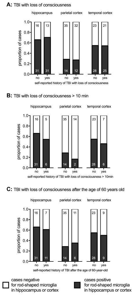
A subtype of microglia is defined by the morphological appearance of the cells as rod shaped. Little is known about this intriguing cell type, as there are only a few case reports describing rod-shaped microglia in the neuropathological literature. Rod-shaped microglia were shown recently to account for a substantial proportion of the microglia cells in the hippocampus of both demented and cognitively intact aged individuals. We hypothesized that aging could be a defining feature in the occurrence of rod-shaped microglia. To test this hypothesis, 2 independent series of autopsy cases (total n = 168 cases), which covered the adult lifespan from 20 to 100+ years old, were included in the study. The presence or absence of rod-shaped microglia was scored on IBA1 immunohistochemically stained slides for the hippocampus and cortex. We found that age was one of the strongest determinants for the presence of rod-shaped microglia in the hippocampus and the cortex. We found no association with the presence of rod-shaped microglia and a self-reported history of a TBI. Alzheimer's disease-related pathology was found to influence the presence of rod-shaped microglia, but only in the parietal cortex and not in the hippocampus or temporal cortex. Future studies are warranted to determine the functional relevance of rod-shaped microglia in supporting the health of neurons in the aged brain, and the signaling processes that regulate the formation of rod-shaped microglia.

Keywords: Aging; Alzheimer's disease; Hippocampus; Microglia activation; Neurodegeneration; Neuroinflammation; Neuropathology; Traumatic brain injury.


Conflict of interest statement

Conflict of Interest: None

Figures

Figure 1
Figure 1. Examples of rod-shaped microglia from the UK series
(A) Numerous rod-shaped microglia are found throughout the CA1 region of the hippocampus of an 86-year-old female. The arrow indicates the area shown at a higher magnification in (B). (C) Long thin rod-shaped microglia, which appear to be surrounding a pyramidal neuron, in the subiculum of a 39-year-old male, who had hypoxia / ischemia pathology, and a pulmonary associated cause of death. (D) Example of rod-shaped microglia seen in the frontal cortex of a 28-year-old female, who died of atherosclerotic disease. (E) A number of long trains of rod-shaped microglia were found in the CA1 region of the hippocampus of a 25-year-old female, who had hypoxia / ischemia pathology, and a pulmonary associated cause of death. Scale bar = 50μm
Figure 2
Figure 2. Association of aging on the presence of rod-shaped microglia
(A) In the hippocampus a significant trend was found for an increasing occurrence of rod-shaped microglia with increasing age (p=0.017, Cochran Armitage Trend Test). (B) A significant increase in the occurrence of rod shaped microglia in the hippocampus was found in those individuals 70-years or older (70+) at the time of death, compared to those individuals 20–69-years old at the time of death (p=0.0033, Fisher Exact test) (C) A similar pattern was found for the frontal cortex when plotted as age by decade (p=0.222, Cochran-Armitage Trend Test), or split into young adults (20–69-years old) and older adults (70+) (p=0.089, Fisher Exact test). The number of cases in each group are shown in the bars.
Figure 3
Figure 3. Regional heterogeneity in the occurrence of rod-shaped microglia in younger adults versus aged adults
Cases were subdivided by which brain region(s) rod-shaped microglia were found, as follows: 1) in both the cortex and the hippocampus, 2) in the hippocampus only, 3) in the cortex only. In younger adults (20–69 years old) rod shaped microglia were found in the hippocampus, or in the frontal cortex, which is in contrast to the older adults (70+ years old) (p=0.047, χ2 test). The number of cases in each group are shown in the bars.
Figure 4
Figure 4. Association of age and sex on the presence of rod-shaped microglia
The presence of rod-shaped microglia was determined in cases from the AI-ACT series. (A) Age at the time of death was found to significantly contribute to an increase percentage of cases positive for the presence of rod-shaped microglia. (B) Sex was also found to effect the odds ratio that rod-shaped microglia would be found. See table 2 for summary or results. The number of cases in each group are shown in the bars.
Figure 5
Figure 5. Association between a history of traumatic brain injury (TBI) and the presence of rod-shaped microglia in AI-ACT series
(A) A self-reported history of at least one TBI with a loss of consciousness of at least a few seconds was not found associated with the presence of rod-shaped microglia. No association with the presence of rod-shaped microglia was found with either: (B) a self-reported history of at least one TBI with a loss of consciousness of more than 10 min; or a first TBI exposure after the age of 60 years old. The number of cases in each group are shown in the bars.

References

    1. Abner EL, Nelson PT, Kryscio RJ, Schmitt FA, Fardo DW, Woltjer RL, Cairns NJ, Yu L, Dodge HH, Xiong C, Masaki K, Tyas SL, Bennett DA, Schneider JA, Arvanitakis Z. Diabetes is associated with cerebrovascular but not Alzheimer’s disease neuropathology. Alzheimers Dement. 2016;12(8):882–9. - PMC - PubMed
    1. Abner EL, Nelson PT, Schmitt FA, Browning SR, Fardo DW, Wan L, Jicha GA, Cooper GE, Smith CD, Caban-Holt AM, Van Eldik LJ, Kryscio RJ. Self-reported head injury and risk of late-life impairment and AD pathology in an AD center cohort. Dement Geriatr Cogn Disord. 2014;37(5–6):294–306. - PMC - PubMed
    1. Ackman JB, Siddiqi F, Walikonis RS, LoTurco JJ. Fusion of microglia with pyramidal neurons after retroviral infection. J Neurosci. 2006;26(44):11413–22. - PMC - PubMed
    1. Bachstetter AD, Morganti JM, Jernberg J, Schlunk A, Mitchell SH, Brewster KW, Hudson CE, Cole MJ, Harrison JK, Bickford PC, Gemma C. Fractalkine and CX3CR1 regulate hippocampal neurogenesis in adult and aged rats. Neurobiol Aging. 2011;32(11):2030–44. - PMC - PubMed
    1. Bachstetter AD, Van Eldik LJ, Schmitt FA, Neltner JH, Ighodaro ET, Webster SJ, Patel E, Abner EL, Kryscio RJ, Nelson PT. Disease-related microglia heterogeneity in the hippocampus of Alzheimer’s disease, dementia with Lewy bodies, and hippocampal sclerosis of aging. Acta Neuropathol Commun. 2015:3. - PMC - PubMed

Publication types