. 2017 May;91(5):1104-1114.
doi: 10.1016/j.kint.2016.10.034. Epub 2017 Jan 25.

Recombinant α-Klotho may be prophylactic and therapeutic for acute to chronic kidney disease progression and uremic cardiomyopathy

Affiliations

Recombinant α-Klotho may be prophylactic and therapeutic for acute to chronic kidney disease progression and uremic cardiomyopathy

Ming Chang Hu et al. Kidney Int. 2017 May.

Abstract

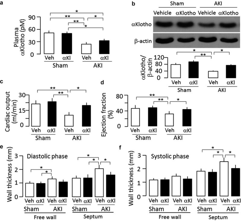
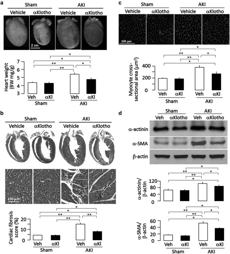
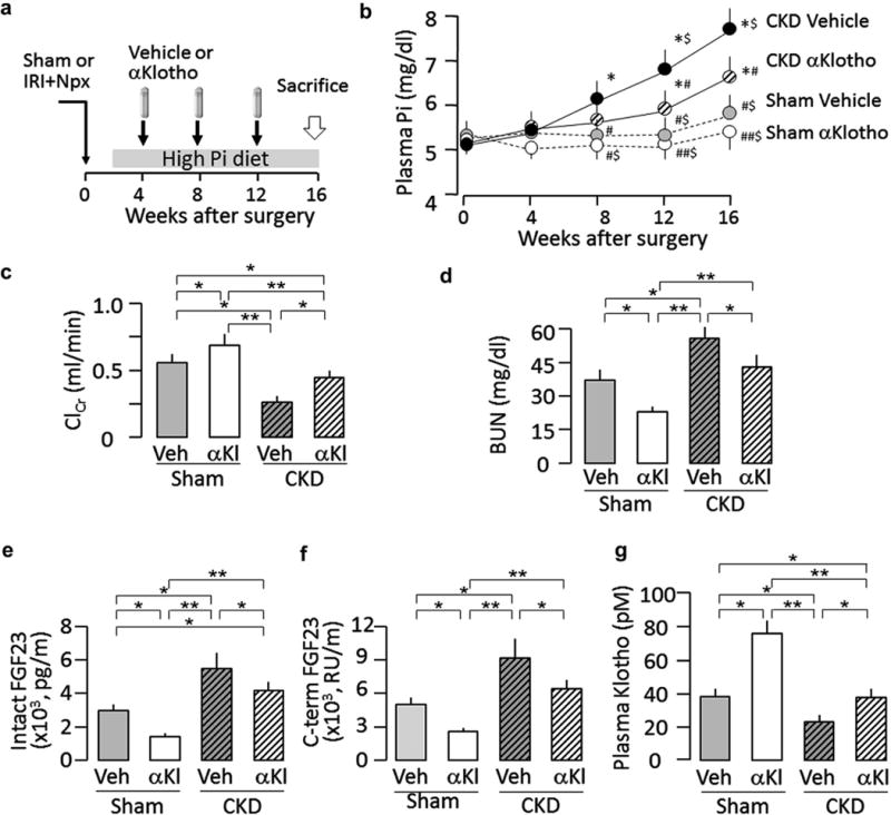
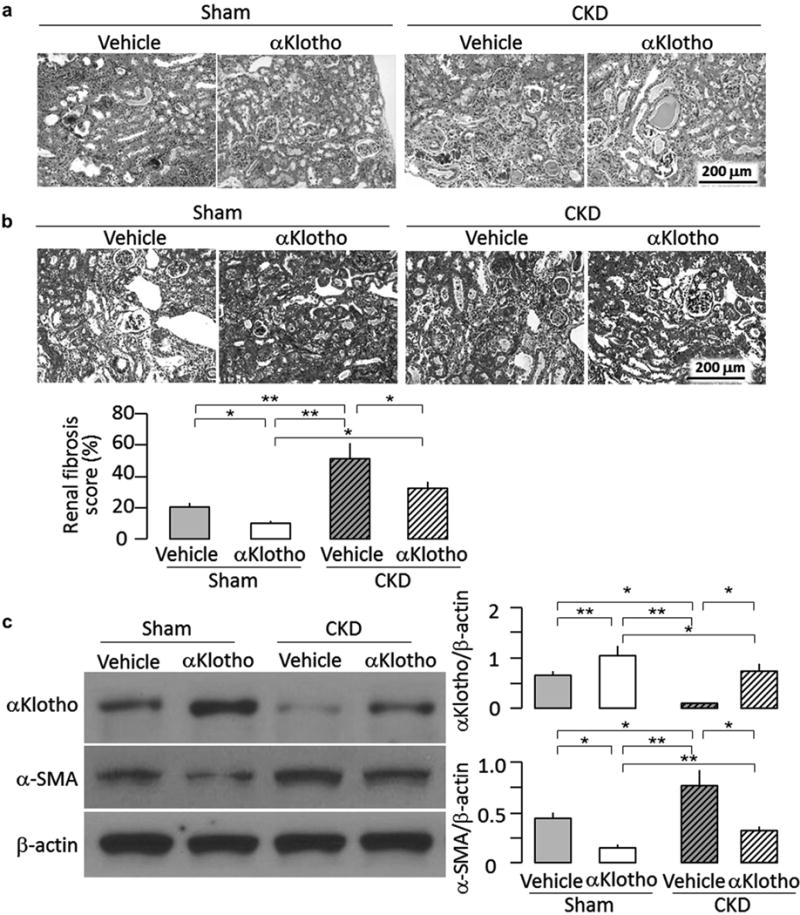
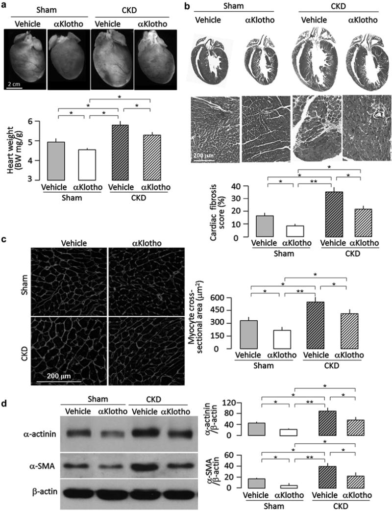
α-Klotho is highly expressed in the kidney, and its extracellular domain is cleaved and released into the circulation. Chronic kidney disease (CKD) is a state of α-Klotho deficiency, which exerts multiple negative systemic effects on numerous organs including the cardiovascular system. Since acute kidney injury (AKI) greatly escalates the risk of CKD development, we explored the effect of α-Klotho on prevention and treatment on post-AKI to CKD progression and cardiovascular disease. Therein, ischemia reperfusion injury-induced AKI was followed by early administration of recombinant α-Klotho or vehicle starting one day and continued for four days after kidney injury (CKD prevention protocol). A CKD model was generated by unilateral nephrectomy plus contralateral ischemia reperfusion injury. Late administration of α-Klotho in this model was started four weeks after injury and sustained for 12 weeks (CKD treatment protocol). The prevention protocol precluded AKI to CKD progression and protected the heart from cardiac remodeling in the post-AKI model. One important effect of exogenous α-Klotho therapy was the restoration of endogenous α-Klotho levels long after the cessation of exogenous α-Klotho therapy. The treatment protocol still effectively improved renal function and attenuated cardiac remodeling in CKD, although these parameters did not completely return to normal. In addition, α-Klotho administration also attenuated high phosphate diet-induced renal and cardiac fibrosis, and improved renal and cardiac function in the absence of pre-existing renal disease. Thus, recombinant α-Klotho protein is safe and efficacious, and might be a promising prophylactic or therapeutic option for prevention or retardation of AKI-to-CKD progression and uremic cardiomyopathy.

Keywords: acute kidney injury; cardiovascular disease; chronic kidney disease; ischemia reperfusion; phosphate.


Conflict of interest statement

DISCLOSURE

MK has a patent on Klotho peptides and antibodies; OM has consulted for AbbVie, Aliena, Ardelyx, Calico, Genzyme-Sanofi, and Takeda. All the other authors declared no competing interests.

Figures

Figure 1
Figure 1. αKlotho administration after acute kidney injury (AKI) maintained higher plasma and renal αKlotho levels and improved cardiac function in ischemia-reperfusion injury (IRI)–induced AKI mice
Sham-treated or IRI-induced AKI wild-type mice were Injected i.p. with αKlotho (αKl) protein (0.01 mg/kg) or vehicle (Veh) (phosphate-buffered saline) for 4 consecutive days starting 24 hours after surgery. Throughout the experimental period, all mice were fed normal rodent chow. At 20 weeks after surgery, animals were killed. (a) Plasma αKlotho. (b) αKlotho protein in the kidney. Upper panel shows representative immunoblots for αKlotho and (β-actin in the kidney. Bottom panel is a summary of normalized protein quantification from all examined immunoblots. (c) Cardiac output, (d) Left ventricular ejection fraction, (e) Left ventricular wall thickness at diastole, (f) Left ventricular wall thickness at systole. Data are expressed as means ± SD of 4 mice from each group, and statistical significance was assessed by 1-way analysis of variance followed by Student-Newman-Keuls test, and accepted when: *P < 0.05, **P < 0.01 between 2 groups.
Figure 2
Figure 2. αKlotho administration attenuated cardiac remodeling after acute kidney injury (AKI)
Wild-type mice subjected to ischemia-reperfusion injury (IRI)–induced AKI and sham surgery were treated with αKlotho (αKl) protein (described in Supplementary Figure S1A) or vehicle (Veh) and killed 20 weeks after surgery. All mice were fed normal rodent chow, (a) Cardiac hypertrophy in mice after AKI. Upper panel shows representative gross macrographs of hearts. Bottom panel is a summary of ratio of heart weight to body weight of examined mice. (b) Cardiac fibrosis in mice after AKI. Upper panel shows representative macrographs of sagittal sections (trichrome stain). Middle panel shows representative micrographs of left ventricular sections (trichrome stain). Bottom panel is a summary of semiquantification of trichrome-positive area to whole heart section calculated using ImageJ software. (c) Hypertrophic cardiomyocytes in post-AKI mice. Upper panel shows representative micrographs of heart sections stained with WGA. Bottom panel is a summary of cardiomyocyte size calculated using ImageJ software, (d) Hypertrophic and fibrotic markers in the heart. Upper panel shows representative immunoblots for α-actinin, α-smooth muscle actin (α-SMA), and (β-actin protein. Bottom panel shows a summary of normalized protein quantification from all examined immunoblots. Data are expressed as means ± SD of 4 mice from each group, and statistical significance was assessed by 1-way analysis of variance followed by Student-Newman-Keuls test and accepted when: *P < 0.05, **P < 0.01 between 2 groups. WGA, wheat germ agglutinin.
Figure 3
Figure 3. Effects of chronic administration of αKlotho (αKl) after established chronic kidney disease (CKD) on plasma phosphate, renal function, plasma FGF23, and plasma αKlotho in CKD mice
(a) Animal experimental design. Using osmotic minipump implants from week 4 to 16 after induction of CKD. The minipumps contained vehicle (Veh; phosphate-buffered saline) or αKlotho (0.3 μg/g body weight). All mice were fed normal rodent chow or high-phosphate (2%) experimental chow for 12 weeks starting 2 weeks after surgery. (b) Plasma phosphate 0,4,8,12, and 16 weeks after surgery. (c) Creatinine clearance (ClCr) at 16 weeks. (d) Blood urea nitrogen (BUN) at 16 weeks. (e) Plasma intact FGF23 at 16 weeks, (f) Plasma C-terminal FGF23 (C-term FGF23) at 16 weeks, (g) Plasma αKlotho at 16 weeks. Data are expressed as means ± SD of 4 mice from each group, and statistical significance was assessed by 1-way analysis of variance followed by Student-Newman-Keuls test and accepted when: *P < 0.05 versus sham-vehicle; #P < 0.05, ##P < 0.01 versus CKD-vehicle; $P < 0.05 versus CKD-αKlotho at 0, 4, 8,12, and 16 weeks of surgery, respectively, for (b). *P < 0.05, **P < 0.01 between 2 groups for (c–g). IRI, ischemia-reperfusion injury; Npx, nephrectomy; Pi, phosphate.
Figure 4
Figure 4. Effects of chronic administration of αKlotho after established chronic kidney disease (CKD) on kidney histology and renal αKlotho expression
Animal experimental design was shown in Figure 3a. At 16 weeks, the kidneys were harvested for renal histology and immunoblot analysis, (a) Representative renal micrographs of hematoxylin-eosin stained kidney sections, (b) Renal fibrosis in mice at 16 weeks. Upper panel shows representative micrographs of trichrome-stained kidney sections. Bottom panel shows renal fibrosis scores from trichrome section calculated using ImageJ software, (c) αKlotho and fibrotic markers in the kidney. Left panel shows representative immunoblots for αKlotho, α-smooth muscle actin (α-SMA), and β-actin protein. Right panel shows a summary of normalized protein quantification from all examined immunoblots. Data are expressed as means ± SD of 4 mice from each group. Statistical significance was assessed by 1-way analysis of variance followed by Student-Newman-Keuls test and accepted when: *P < 0.05, **P < 0.01 between 2 groups.
Figure 5
Figure 5. Effects of chronic administration of αKlotho on cardiac remodeling in chronic kidney disease (CKD) mice
Animal experimental design was shown in Figure 3a. At 16 weeks after surgery, hearts were harvested for histology, immunoblot, and Immunohlstochemlstry. (a) Cardiac hypertrophy In CKD mice. Upper panel shows representative gross macrographs of hearts. Bottom panel shows a summary of ratio of heart weight to body weight for the examined mice, (b) Cardiac fibrosis in CKD mice. Upper panel shows representative macrographs of sagittal sections (trichrome stain). Middle panel shows representative micrographs of left ventricular sections (trichrome stain). Bottom panel is a summary of semiquantification of trichrome-positive area to whole heart section performed using ImageJ software. Data are expressed as means ± SD of 4 mice from each group, (c) Hypertrophic cardiomyocytes in CKD mice. Left panel shows representative micrographs of left ventricular sections stained with wheat germ agglutinin. Right panel is a summary of cardiomyocyte size calculated using ImageJ software, (d) Hypertrophic and fibrotic markers in the heart. Left panel shows representative immunoblotsfor α-actinin, α-smooth muscle actin (α-SMA), and β-actin protein. Right panel is a summary of normalized protein quantification from all examined immunoblots. Data are expressed as means ± SD of 4 mice from each group. Statistical significance was assessed by 1-way analysis of variance followed by Student-Newman-Keuls test and accepted when: *P < 0.05, **P < 0.01 between 2 groups.

References

    1. Kuro-o M, Matsumura Y, Aizawa H, et al. Mutation of the mouse klotho gene leads to a syndrome resembling ageing. Nature. 1997;390:45–51. - PubMed
    1. Kurosu H, Yamamoto M, Clark JD, et al. Suppression of aging in mice by the hormone Klotho. Science. 2005;309:1829–1833. - PMC - PubMed
    1. Hu MC, Shi M, Zhang J, et al. Renal production, uptake, and handling of circulating alphaKlotho. J Am Soc Nephrol. 2016;27:79–90. - PMC - PubMed
    1. Chen CD, Tung TY, Liang J, et al. Identification of cleavage sites leading to the shed form of the anti-aging protein klotho. Biochemistry. 2014;53:5579–5587. - PMC - PubMed
    1. van Loon EP, Pulskens WP, van der Hagen EA, et al. Shedding of klotho by ADAMs in the kidney. Am J Physiol Renal Physiol. 2015;309:F359–F368. - PubMed

MeSH terms