. 2017 Mar 27:346:226-237.
doi: 10.1016/j.neuroscience.2017.01.025. Epub 2017 Jan 25.

Involvement of PKCα and ERK1/2 signaling pathways in EGCG's protection against stress-induced neural injuries in Wistar rats

Affiliations

Involvement of PKCα and ERK1/2 signaling pathways in EGCG's protection against stress-induced neural injuries in Wistar rats

Xiaoling Zhao et al. Neuroscience. .

Abstract

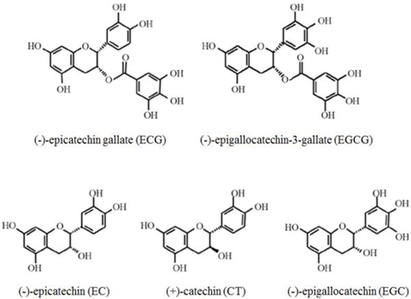
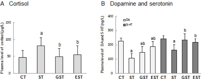
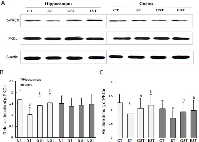
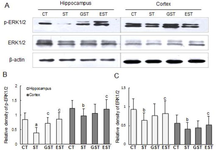
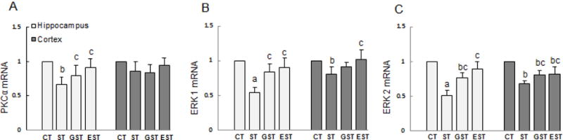
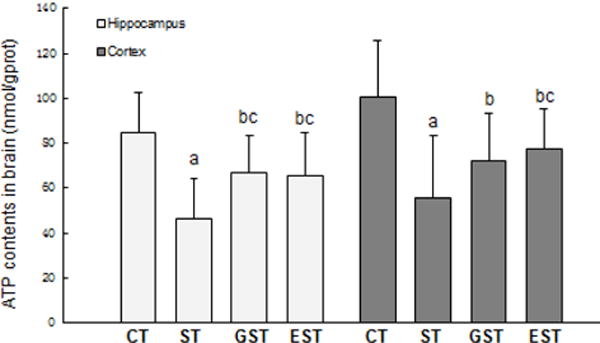
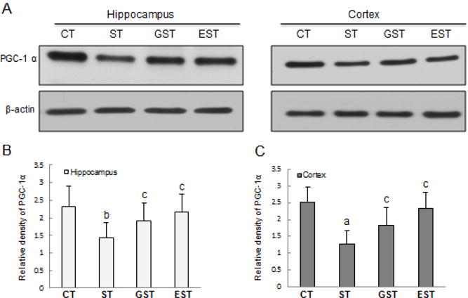
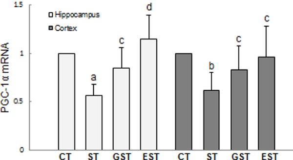
Stress-induced neural injuries are closely linked to the pathogenesis of various neuropsychiatric disorders and psychosomatic diseases. We and others have previously demonstrated certain protective effects of epigallocatechin-3-gallate (EGCG) in stress-induced cerebral impairments, but the underlying protective mechanisms still remain poorly elucidated. Here we provide evidence to support the possible involvement of PKCα and extracellular signal-regulated kinase 1/2 (ERK1/2) signaling pathways in EGCG-mediated protection against restraint stress-induced neural injuries in rats. In both open-field and step-through behavioral tests, the restraint stress-induced neuronal impairments were significantly ameliorated by administration of EGCG or green tea polyphenols (GTPs), which was associated with a partial restoration of normal plasma glucocorticoid, dopamine and serotonin levels. Furthermore, the stress-induced decrease of PKCα and ERK1/2 expression and phosphorylation was significantly attenuated by EGCG and to a less extent by GTP administration. Additionally, EGCG supplementation restored the production of adenosine triphosphate (ATP) and the expression of a key regulator of cellular energy metabolism, the peroxisome proliferators-activated receptor-γ coactivator-1α (PGC-1α), in stressed animals. In conclusion, PKCα and ERK1/2 signaling pathways as well as PGC-1α-mediated ATP production might be involved in EGCG-mediated protection against stress-induced neural injuries.

Keywords: epigallocatechin-3-gallate (EGCG); extracellular signal-regulated kinase1/2 (ERK1/2); peroxisome proliferators-activated receptor-γ coactivator-1 α (PGC-1α); protein kinase C α (PKCα); stress.


Conflict of interest statement

The authors declare no conflict of interest.

Figures

Figure 1
Figure 1. The chemical structures of Green tea polyphenols (GTPs) (Goodman et al. 2013)
Based on the structural variations, GTPs are categorized into 5 derivatives, including epicatechin gallate (ECG), epigallocatechin-3-gallate (EGCG), epicatechin (EC), catechin (CT) and epigallocatechin (EGC).
Figure 2
Figure 2. The levels of plasma cortisol, dopamine and serotonin
Wistar rats weighing 140–160g were randomly assigned into 4 groups (10 animals per group): normal control (CT), stress control (ST), stress and GTPs-modulation (GST), as well as stress and EGCG-modulation (EST) groups. Stress rats received restraint for 4 weeks; GTPs and EGCG were given to animals by intragastric administration once a day and for 4 weeks. The whole blood samples were collected immediately after the rats were sacrificed, and were centrifuged to separate plasma. The contents of cortisol, dopamine (DA) and serotonin (5-HT) were measured, and expressed as mean ± SD. a P<0.05 vs CT group;b P<0.05 vs ST group
Figure 3
Figure 3. Expression and phosphorylation of PKCα in the hippocampus and cortex of rats
The hippocampus and cortex samples were quickly harvested after the animals were sacrificed, and snap-frozen for tissue homogenization. Proteins (50–100 μg) from tissue lysates were resolved by SDS-PAGE, and blotted with specific antibodies against PKC α or p-PKC α. The specific signals were visualized with “ChemiDoc XRS’’ digital imaging system, and a representative Western blot was shown. β-actin was used as an internal control(A). The relative protein levels were expressed as the relative band density of the corresponding protein (B, C). a P<0.05 vs CT group;b P<0.05 vs ST group
Figure 4
Figure 4. Expressions of ERK1/2 and phosphorylated ERK1/2 in the hippocampus and cortex of rats
Proteins (50–100 μg) of the hippocampus and cortex tissue lysates were resolved by SDS-PAGE, and blotted with specific antibodies against ERK1/2 or p-ERK1/2. The specific signals were visualized with “ChemiDoc XRS’’ digital imaging system, and a representative Western blot was shown. β-actin was used as an internal control(A). The relative protein levels were expressed as the relative band density of the corresponding protein (B, C). a P<0.01, b P<0.05 vs CT group;c P<0.05 vs ST group
Figure 5
Figure 5. The expressions of PKCα mRNA and ERK1/2 mRNA in the hippocampus and cortex of rats
The samples of the hippocampus and cortex were quickly isolated after the animals were sacrificed, and snap-frozen for tissue homogenization. Total RNA was isolated, and the expression levels of PKCα and ERK1/2 mRNA were determined by real-time RT-PCR, and expressed as mean ± SD of Gapdh mRNA levels. a P<0.01, b P<0.05 vs CT group;c P<0.05 vs ST group.
Figure 6
Figure 6. The productions of ATP in the brain of rats
The samples of the hippocampus and cortex were quickly isolated after the animals were sacrificed, and snap-frozen for tissue homogenization. The tissue levels of ATP was measured, and expressed as mean ± SD. a P<0.01, b P<0.05 vs CT group;c P<0.05 vs ST group.
Figure 7
Figure 7. The expression of PGC-1α in the hippocampus and cortex of rats
The hippocampus and cortex samples were quickly harvested after the animals were sacrificed, and snap-frozen for tissue homogenization. Proteins (50–100 μg) from tissue lysates were resolved by SDS-PAGE, and blotted with specific antibody against PGC-1 α, and the specific signals were visualized with “ChemiDoc XRS’’ digital imaging system. β-actin was used as an internal control. A representative Western blot result was shown (A). The quantitation of protein bands is expressed as relative density of the corresponding protein (B, C). a P<0.01, b P<0.05 vs CT group;c P<0.05 vs ST group.
Figure 8
Figure 8. The expression of PGC-1α mRNA in the hippocampus and cortex of rats
The hippocampus and cortex samples were quickly isolated after the animals were sacrificed, and snap-frozen for tissue homogenization. Total RNA was isolated, the expression of PGC-1α mRNA was determined by real-time RT-PCR, and expressed as mean ± SD of Gapdh mRNA levels. a P<0.01, b P<0.05 vs CT group;c P<0.05, d P<0.01 vs ST group.

References

    1. Abd El Mohsen MM, Kuhnle G, Rechner AR, Schroeter H, Rose S, Jenner P, Rice-Evans CA. Uptake and metabolism of epicatechin and its access to the brain after oral ingestion. Free Radic Biol Med. 2002;33:1693–1702. - PubMed
    1. Adams F, Grassie M, Shahid M, Hill DR, Henry B. Acute oral dexamethasone administration reduces levels of orphan GPCR glucocorticoid-induced receptor (GIR) mRNA in rodent brain: potential role in HPA-axis function. Brain Res Mol Brain Res. 2003;117:39–46. - PubMed
    1. Angelucci L. The glucocorticoid hormone: from pedestal to dust and back. Eur J Pharmcol. 2000;405:139–147. - PubMed
    1. Auger C, Mullen W, Hara Y, Crozier A. Bioavailability of polyphenon E flavan-3-ols in humans with an ileostomy. J Nutr. 2008;138:1535S–1542S. - PubMed
    1. Baptista FI, Henriques AG, Silva AM, Wiltfang J, da Cruze Silva OA. Flavonoids as therapeutic compounds targeting key proteins involved in Alzheimer’s disease. ACS Chem Neurosci. 2014;5:83–92. - PMC - PubMed

Publication types

MeSH terms