

Review
doi: 10.1016/j.tcb.2016.12.002.
Epub 2017 Feb 9.
New Insights into the Physiological Role of Endoplasmic Reticulum-Associated Degradation
Affiliations
- PMID: 28131647
- PMCID: PMC5440201
- DOI: 10.1016/j.tcb.2016.12.002
Item in Clipboard
Review
New Insights into the Physiological Role of Endoplasmic Reticulum-Associated Degradation
Trends Cell Biol.
2017 Jun.
Abstract
Many human diseases are associated with mutations causing protein misfolding and aggregation in the endoplasmic reticulum (ER). ER-associated degradation (ERAD) is a principal quality-control mechanism responsible for targeting misfolded ER proteins for cytosolic degradation. However, despite years of effort, the physiological role of ERAD in vivo remains largely unknown. Several recent studies have reported intriguing phenotypes of mice deficient for ERAD function in specific cell types. These studies highlight that mammalian ERAD has been designed to perform a wide-range of cell-type-specific functions in vivo in a substrate-dependent manner.
Copyright © 2016 Elsevier Ltd. All rights reserved.
Figures
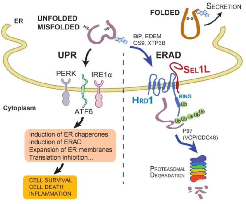
While folded proteins exit the ER, terminally unfolded or misfolded proteins in the ER activate UPR via three sensors IRE1α, PERK and ATF6, which initiate multiple signaling pathways leading to the induction of ER chaperones and ERAD, expansion of ER membranes and translation inhibition. In addition, misfolded proteins can be recruited to the ERAD complex via the activity of various ER chaperones such as BiP, EDEM, OS9 and XTP3B for cytosolic degradation. The Sel1L-Hrd1 protein complex represents the most conserved ERAD complex in mammals. Following retrotranslocation into the cytosol, substrates are ubiquitinated and, with the help of p97 (VCP/CDC48), degraded by the proteasome in the cytosol.
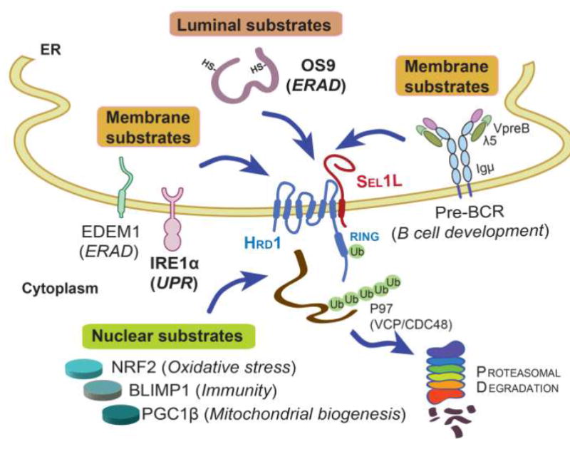
Three types of misfolded proteins, ER luminal (OS9), transmembrane (EDEM1, IRE1α and pre-BCR), and nuclear proteins (NRF2, PGC1β and BLIMP1), have been identified as Sel1L-Hrd1 ERAD substrates using ERAD-deficient mouse models. Substrates in bold (OS9 and IRE1α) have been tested in several different cell types while those in plain fonts have been tested in only one cell type. Substrate-related function is indicated in parentheses. Among all the substrates, the pre-BCR protein complex is unique in that it is consisted of heavy chain Igµ and light chains λ5 and VpreB.
References
Publication types
MeSH terms
Grants and funding
LinkOut - more resources
Full Text Sources
Other Literature Sources
