Review
. 2016;62(3):335-342.

Structural insights into cell cycle control by essential GTPase Era

Affiliations
Review

Structural insights into cell cycle control by essential GTPase Era

Xinhua Ji. Postepy Biochem. 2016.

Abstract

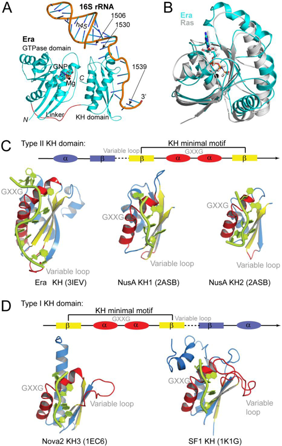
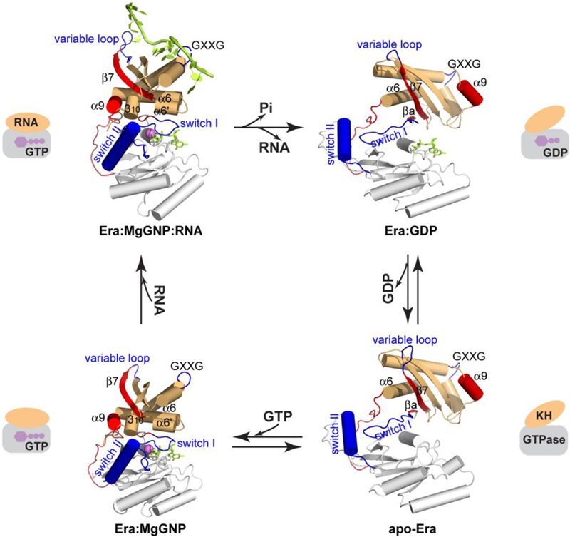
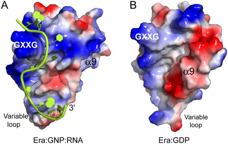
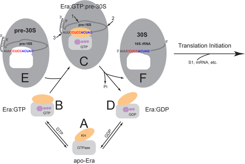
Era (Escherichia coli Ras-like protein), essential for bacterial cell viability, is composed of an N-terminal GTPase domain and a C-terminal KH domain. In bacteria, it is required for the processing of 16S ribosomal RNA (rRNA) and maturation of 30S (small) ribosomal subunit. Era recognizes 10 nucleotides (1530GAUCACCUCC1539) near the 3' end of 16S rRNA and interacts with helix 45 (h45, nucleotides 1506-1529). GTP binding enables Era to bind RNA, RNA binding stimulates Era's GTP-hydrolyzing activity, and GTP hydrolysis releases Era from matured 30S ribosomal subunit. As such, Era controls cell growth rate via regulating the maturation of the 30S ribosomal subunit. Ribosomes manufacture proteins in all living organisms. The GAUCA sequence and h45 are highly conserved in all three kingdoms of life. Homologues of Era are present in eukaryotic cells. Hence, the mechanism of bacterial Era action also sheds light on the cell cycle control of eukaryotes.

Białko Era (ang. Escherichia coli Ras-like protein), niezbędne dla funkcjonowania komórki bakteryjnej, składa się z N-końcowej domeny o aktywności GTPazy oraz C-końcowej domeny KH. W komórce bakteryjnej białko to uczestniczy w modyfikacji rybosomalnego RNA (rRNA) 16S oraz dojrzewaniu podjednostki 30S (małej) rybosomu. Era rozpoznaje sekwencję 10 nukleotydów (1530GAUCACCUCC1539) w pobliżu końca 3' rRNA 16S i oddziałuje z helisą 45 (h45, nukleotydy 1506-1529). Związanie GTP umożliwia związanie przez białko Era cząsteczki RNA, co zwiększa jego aktywność hydrolizy GTP. W wyniku hydrolizy GTP białko Era zostaje z kolei uwolnione od dojrzałej podjednostki 30S rybosomu. W ten sposób Era kontroluje szybkość wzrostu komórki poprzez regulację dojrzewania podjednostek 30S rybosomów. Jak wiadomo, rybosomy są miejscem produkcji białek w każdej komórce. Sekwencja GAUCA i struktura h45 są zachowane w ewolucji we wszystkich trzech królestwach organizmów żywych. Homologi białka Era wykryto również w komórkach eukariotycznych. Z tego względu zrozumienie mechanizmu działania bakteryjnego białka Era dostarcza ważnych wskazówek dotyczących kontroli cyklu komórkowego także w komórkach Eukaryota.

Keywords: 16S rRNA; 30S ribosomal subunit; cell cycle control; ribosome biogenesis.


Figures

Figure 1
Figure 1
Overall structure of Era. (A) Schematic representation of the Era:MgGNP:RNA301 complex embedded in the Era:MgGNP:RNA301:KsgA structure (PEB entry 3R9X), showing the GTPase domain, KH domain, Mg2+ ion, non-hydrolysable GTP-analog GNP, and RNA301 that contains helix 45 (h45) and the 3’ tail of 16S rRNA. The protein is illustrated as a ribbon diagram (spirals, helices; arrows, strands; and tubes, loops), the Mg2+ ion as a sphere, the GNP molecule as a stick model, and the RNA as a cartoon. Atomic color scheme is used (N, blue; C, cyan; O, red; phosphorus, orange; and Mg, black) except that the linker between the two domains of Era is highlighted in red. (B) The GTPase domain and MgGNP of the Era:MgGNP:RNA301 structure are superimposed with the Ras:MgGNP structure (in grey, PDB entry 4RSG). (C) Type II KH:RNA complexes in the Era:MgGNP:RNA (PDB entry 3IEV) and NusA-RNA (PDB entry 2ASB) structures. (D) Type I KH:RNA complexes in the Nova2:RNA (PDB entry 1EC6) and SF1:RNA (PDB entry 1K1G) structures. The KH domains are aligned on the basis of the helix-turn-helix (HTH) motif, which contains the GXXG loop, and shown separately for clarity. Nucleotide residues that are recognized by the KH domains are 1531AUCACCUCC1539 for Era-KH, 42AGAA45 for NusA-KH1, 48CAAUA52 for NusA-KH2, 12UCAC15 for Nova2, and 6UAAC9 for SF1. Linear representations are used to illustrate the topological differences between the two types of KH folds, in which the dashed line represents the variable loop and the GXXG represents the GXXG loop in the HTH motif. Panels C and D were originally published in Reference [12].
Figure 2
Figure 2
The functional cycle of Era as an essential GTPase. The crystal structures of Era are shown based on superimposed GTPase domains, including Era:GNP:RNA (PDB entry 3IEV) and Era:GNP (PDB entry 1WF3) sharing the closed conformation, and apo-Era (PDB entry 1EGA) and Era:GDP (PDB entry 3IEU) sharing the open conformation. The GTPase domain, the linker and the KH domain are shown in white, red and orange, respectively. Switches I and II and the GXXG and variable loops are highlighted in blue, and the α9 helix, βa and β7 strands, and linker are highlighted in red. The GNP and RNA molecules are colored in green, and the Mg2+ ion in magenta. Highly simplified representation of each structure is composed of a rectangle and an ellipse. This figure was adopted from Reference [12] with revisions and additions.
Figure 3
Figure 3
Structure-based sequence alignment of Era proteins from A. aeolicus, T. thermophilus, and E. coli (PDB entries 3IEV, 1WF3, and 1EGA, respectively). Secondary structural elements and the position of the inter-domain 17-residue linker are indicated above the sequences. Switch regions I and II and the GXXG and variable loops are indicated. Identical residues and similar residues are shaded in dark and light green, respectively.
Figure 4
Figure 4
Role of α9 in the regulation of RNA binding by KH. (A) Electrostatic-surface representation of the KH domain and RNA in the Era:GNP:RNA structure (PDB entry 3IEV). (B) Electrostatic-surface representation of the KH domain in the Era:GDP structure (PDB entry 3IEU). On the molecular surfaces, positively charged areas are indicated in blue and negatively charged areas in red. The RNA is illustrated as a cartoon in green. The GXXG motif, variable loop, and α9 helix are indicated. This figure was originally published in Reference [12].
Figure 5
Figure 5
GTP-hydrolyzing activity of Era. (A) GTPase activity of Era in the absence of RNA, in the presence of the 1531AUCACCUCCUUA1542 sequence, in the presence of the sequence with 1531–1534 mutations, and in the presence of the sequence with 1535–1539 mutations. (B) GTPase activity of Era in the presence of the 1531AUCACCUCCUUA1542 sequence with a single mutation at each position from 1531 to 1539 or the deletion of nucleotide 1531. (C) GTPase activity of Era in the presence of the 1530GAUCACCUCCUUA1542 sequence, in the presence of the sequence with a single or double mutation at 1530 and/or 1531, or with the deletion of nucleotide 1530 and a single mutation at 1531. (D) GTPase activity of Era in the presence of longer RNA consisted of h45 (nucleotides 1506–1529) and the 1530GAUCACCUCCUUA1542 sequence, either native (RNA301) or with a G1530A mutation. Data were taken from References [12, 13] and presented graphically here.
Figure 6
Figure 6
The recognition of G1530 by Era. (A) Details of Era-G1530 recognition. G1530 and the side chains of E208, E209, R241, and N243 are illustrated as ball-and-stick models in atomic colors (C in green or yellow, O in red, N in blue, and P in orange). Dashed lines in black indicate hydrogen bonds and their distances are in Å. (B) Model of the G1530A mutant, suggesting that the mutant is not favorable for the recognition by Era due to the loss of two hydrogen bonds (between E208/E209 and the Gua base, panel A) and the creation of a repulsive interaction (between R241 and the Ade base, indicated with a red cross). (C) Models of the E209A, E209K, and E209Q mutants. Indicated is the impact of each mutation on the pseudo-base paring interaction between the G1530 base and the side chains of residues 209 and 241. (D) The E209Q mutant of Era is cold sensitive. Compared with the wild type (W3110, on the left of each sub-panel), the E209Q mutant (on the right of each sub-panel) grows slower at all temperatures tested. Growth impairment is observed at 37 and 42°C. At 32°C, small colonies appear after three days (3d). At 25°C, however, colonies do not appear even after 14 days (14d). This figure was originally published in Reference [13].
Figure 7
Figure 7
The recognition of nucleotide residues A1531 and A1534 by Era in stereo. (A) Interactions between A1531 and Era. (B) Interactions between A1534 and Era. Protein and nucleotide residues are shown as stick models in atomic colors (C, green; N, blue; O, red; and P, orange). Protein backbone is shown as a cartoon (helices as spirals, strands as arrows, and loops as tubes), and protein surface is shown with 50% transparency. This figure was originally published in Reference [13].
Figure 8
Figure 8
The functional cycle of Era. As also shown in Figure 2, the GTPase domain is represented by a grey rectangle, the KH domain by an orange oval, and the GTP and GDP molecules by purple cartoons. The pre-30S particle and 30S ribosomal subunit are represented by larger grey ovals. The pre-16S rRNA (an RNase III cleavage product with a 26-bp stem and a 2-nt 3′ overhang) and 16S rRNA are represented by a grey line with embedded 1530GAUCACCUCCUUA1542 sequence at the 3′ end. The highly conserved GAUCA sequence is colored in blue. The anti-Shine-Dalgarno (anti-SD) sequence CCUCC, which is unique for bacteria, is in red. The unoccupied Era-binding pocket in the pre-30S particle and that in the 30S ribosomal subunit are indicated in white. Note that the shapes of the pocket are different in the pre-30S particle and the mature 30S subunit. The four functional states, including (A) apo-Era, (B) Era:GTP, (C) Era:GTP:pre-30S and (D) Era:GDP, are represented by the apo-Era (PDB entry 1EGA), Era:GNP (PDB entry 1WF3), Era:GNP:RNA (PDB entry 3IEV), and Era:GDP (PDB entry 3IEU) structures. In panel C, the cleavage sites of RNase E, RNase G and the unknown nuclease are indicated with numbered arrows 1, 2 and 3, respectively. (E) The pre-30S particle contains pre-16S rRNA. (F) The mature 30S ribosomal subunit contains 16S rRNA. This figure was adopted from Reference [12] with updates.

Similar articles

Cited by

References

    1. Ahnn J, March PE, Takiff HE, Inouye M (1986) A GTP-binding protein of Escherichia coli has homology to yeast RAS proteins. Proc Natl Acad Sci USA 83: 8849–8853 - PMC - PubMed
    1. March PE, Lerner CG, Ahnn J, Cui X, Inouye M (1988) The Escherichia coli Ras-like protein (Era) has GTPase activity and is essential for cell growth. Oncogene 2: 539–544 - PubMed
    1. Inada T, Kawakami K, Chen SM, Takiff HE, Court DL, Nakamura Y (1989) Temperature-sensitive lethal mutant of ERA, a G protein in Escherichia coli. Journal of Bacteriology 171: 5017–5024 - PMC - PubMed
    1. Zuber M, Hoover TA, Dertzbaugh MT, Court DL (1997) A Francisella tularensis DNA clone complements Escherichia coli defective for the production of Era, an essential Ras-like GTP-binding protein. Gene 189: 31–34 - PubMed
    1. Eltsov M, Zuber B (2006) Transmission electron microscopy of the bacterial nucleoid. J Struct Biol 156: 246–254 - PubMed

MeSH terms

LinkOut - more resources