

Biallelic Mutation of ARHGEF18, Involved in the Determination of Epithelial Apicobasal Polarity, Causes Adult-Onset Retinal Degeneration
- PMID: 28132693
- PMCID: PMC5294887
- DOI: 10.1016/j.ajhg.2016.12.014
Biallelic Mutation of ARHGEF18, Involved in the Determination of Epithelial Apicobasal Polarity, Causes Adult-Onset Retinal Degeneration
Abstract
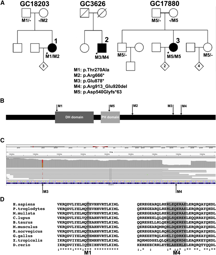
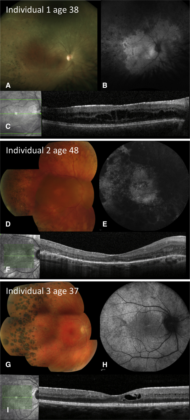
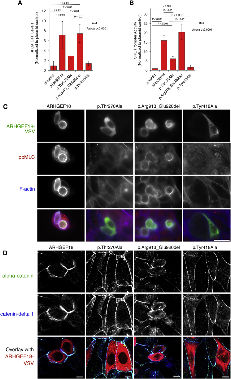
Mutations in more than 250 genes are implicated in inherited retinal dystrophy; the encoded proteins are involved in a broad spectrum of pathways. The presence of unsolved families after highly parallel sequencing strategies suggests that further genes remain to be identified. Whole-exome and -genome sequencing studies employed here in large cohorts of affected individuals revealed biallelic mutations in ARHGEF18 in three such individuals. ARHGEF18 encodes ARHGEF18, a guanine nucleotide exchange factor that activates RHOA, a small GTPase protein that is a key component of tight junctions and adherens junctions. This biological pathway is known to be important for retinal development and function, as mutation of CRB1, encoding another component, causes retinal dystrophy. The retinal structure in individuals with ARHGEF18 mutations resembled that seen in subjects with CRB1 mutations. Five mutations were found on six alleles in the three individuals: c.808A>G (p.Thr270Ala), c.1617+5G>A (p.Asp540Glyfs∗63), c.1996C>T (p.Arg666∗), c.2632G>T (p.Glu878∗), and c.2738_2761del (p.Arg913_Glu920del). Functional tests suggest that each disease genotype might retain some ARHGEF18 activity, such that the phenotype described here is not the consequence of nullizygosity. In particular, the p.Thr270Ala missense variant affects a highly conserved residue in the DBL homology domain, which is required for the interaction and activation of RHOA. Previously, knock-out of Arhgef18 in the medaka fish has been shown to cause larval lethality which is preceded by retinal defects that resemble those seen in zebrafish Crumbs complex knock-outs. The findings described here emphasize the peculiar sensitivity of the retina to perturbations of this pathway, which is highlighted as a target for potential therapeutic strategies.
Keywords: ARHGEF18; apicobasal polarity; inherited retinal dystrophy; p114RhoGEF; retinal degeneration; retinitis pigmentosa.
Copyright © 2017 American Society of Human Genetics. Published by Elsevier Inc. All rights reserved.
Figures
Similar articles
-
ArhGEF18 regulates RhoA-Rock2 signaling to maintain neuro-epithelial apico-basal polarity and proliferation.Development. 2013 Jul;140(13):2787-97. doi: 10.1242/dev.096487. Epub 2013 May 22. Development. 2013. PMID: 23698346
-
Identification of Arhgef12 and Prkci as genetic modifiers of retinal dysplasia in the Crb1rd8 mouse model.PLoS Genet. 2022 Jun 8;18(6):e1009798. doi: 10.1371/journal.pgen.1009798. eCollection 2022 Jun. PLoS Genet. 2022. PMID: 35675330 Free PMC article.
-
The correlation between CRB1 variants and the clinical severity of Brazilian patients with different inherited retinal dystrophy phenotypes.Sci Rep. 2017 Aug 17;7(1):8654. doi: 10.1038/s41598-017-09035-1. Sci Rep. 2017. PMID: 28819299 Free PMC article.
-
Towards understanding CRUMBS function in retinal dystrophies.Hum Mol Genet. 2006 Oct 15;15 Spec No 2:R235-43. doi: 10.1093/hmg/ddl195. Hum Mol Genet. 2006. PMID: 16987889 Review.
-
CRB1 mutations in inherited retinal dystrophies.Hum Mutat. 2012 Feb;33(2):306-15. doi: 10.1002/humu.21653. Epub 2011 Dec 27. Hum Mutat. 2012. PMID: 22065545 Free PMC article. Review.
Cited by
-
δ-Tocotrienol feeding modulates gene expression of EIF2, mTOR, protein ubiquitination through multiple-signaling pathways in chronic hepatitis C patients.Lipids Health Dis. 2018 Jul 21;17(1):167. doi: 10.1186/s12944-018-0804-7. Lipids Health Dis. 2018. PMID: 30031388 Free PMC article.
-
Establishment and functional studies of a model of cardiomyopathy with cardiomyocyte-specific conditional knockout of Arhgef18.Dis Model Mech. 2025 Mar 1;18(3):dmm052172. doi: 10.1242/dmm.052172. Epub 2025 Mar 31. Dis Model Mech. 2025. PMID: 40159883 Free PMC article.
-
Establishment and identification of cardiomyocyte arhGEF18 gene conditional knockout mice.Pediatr Discov. 2023 Aug 14;1(2):e20. doi: 10.1002/pdi3.20. eCollection 2023 Sep. Pediatr Discov. 2023. PMID: 40625715 Free PMC article.
-
EYS is a major gene involved in retinitis pigmentosa in Japan: genetic landscapes revealed by stepwise genetic screening.Sci Rep. 2020 Nov 27;10(1):20770. doi: 10.1038/s41598-020-77558-1. Sci Rep. 2020. PMID: 33247286 Free PMC article.
-
Genetic dissection of non-syndromic retinitis pigmentosa.Indian J Ophthalmol. 2022 Jul;70(7):2355-2385. doi: 10.4103/ijo.IJO_46_22. Indian J Ophthalmol. 2022. PMID: 35791117 Free PMC article. Review.
References
-
- Niu J., Profirovic J., Pan H., Vaiskunaite R., Voyno-Yasenetskaya T. G Protein betagamma subunits stimulate p114RhoGEF, a guanine nucleotide exchange factor for RhoA and Rac1: regulation of cell shape and reactive oxygen species production. Circ. Res. 2003;93:848–856. - PubMed
-
- Arno G., Hull S., Carss K., Dev-Borman A., Chakarova C., Bujakowska K., van den Born I., Robson A.G., Holder G.E., Michaelides M. Reevaluation of the retinal dystrophy due to recessive alleles of RGR with the discovery of a cis-acting mutation in CDHR1. Invest. Ophthalmol. Vis. Sci. 2016;57:4806–4813. - PubMed
-
- Henderson R.H., Mackay D.S., Li Z., Moradi P., Sergouniotis P., Russell-Eggitt I., Thompson D.A., Robson A.G., Holder G.E., Webster A.R., Moore A.T. Phenotypic variability in patients with retinal dystrophies due to mutations in CRB1. Br. J. Ophthalmol. 2011;95:811–817. - PubMed
-
- Arno G., Hull S., Robson A.G., Holder G.E., Cheetham M.E., Webster A.R., Plagnol V., Moore A.T. Lack of interphotoreceptor retinoid binding protein, caused by homozygous mutation of RBP3, is associated with high myopia and retinal dystrophy. Invest. Ophthalmol. Vis. Sci. 2015;56:2358–2365. - PubMed
Publication types
MeSH terms
Substances
Supplementary concepts
Grants and funding
- MR/K023489/1/MRC_/Medical Research Council/United Kingdom
- G0900098/MRC_/Medical Research Council/United Kingdom
- BB/N014855/1/BB_/Biotechnology and Biological Sciences Research Council/United Kingdom
- MC_PC_14089/MRC_/Medical Research Council/United Kingdom
- MR/M009203/1/MRC_/Medical Research Council/United Kingdom
- MC_EX_MR/M009203/1/MRC_/Medical Research Council/United Kingdom
- MR/K006584/1/MRC_/Medical Research Council/United Kingdom
- G0600237/MRC_/Medical Research Council/United Kingdom
- BB/L007584/1/BB_/Biotechnology and Biological Sciences Research Council/United Kingdom
- 205041/Z/16/Z/WT_/Wellcome Trust/United Kingdom
LinkOut - more resources
Full Text Sources
Other Literature Sources
Molecular Biology Databases
