Comparative Study
. 2017 Feb 16;65(4):671-684.e5.
doi: 10.1016/j.molcel.2016.12.016. Epub 2017 Jan 26.

DNA Double-Strand Break Resection Occurs during Non-homologous End Joining in G1 but Is Distinct from Resection during Homologous Recombination

Affiliations
Comparative Study

DNA Double-Strand Break Resection Occurs during Non-homologous End Joining in G1 but Is Distinct from Resection during Homologous Recombination

Ronja Biehs et al. Mol Cell. .

Abstract

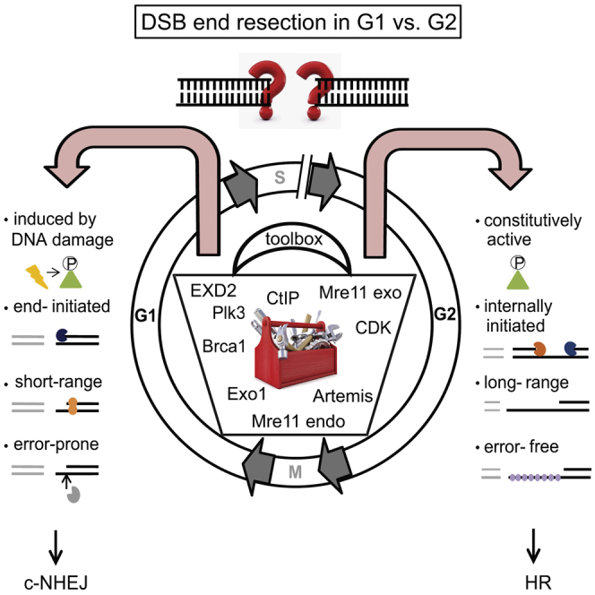
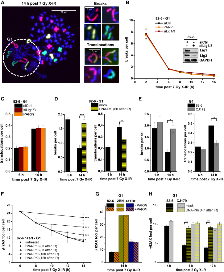
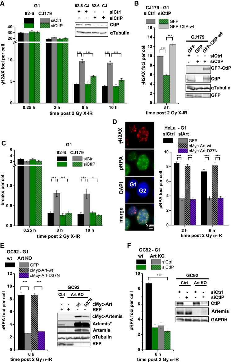
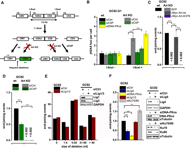
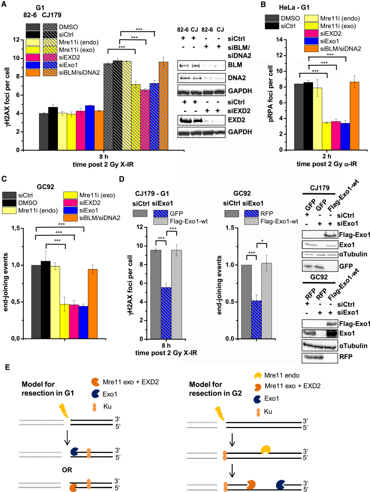
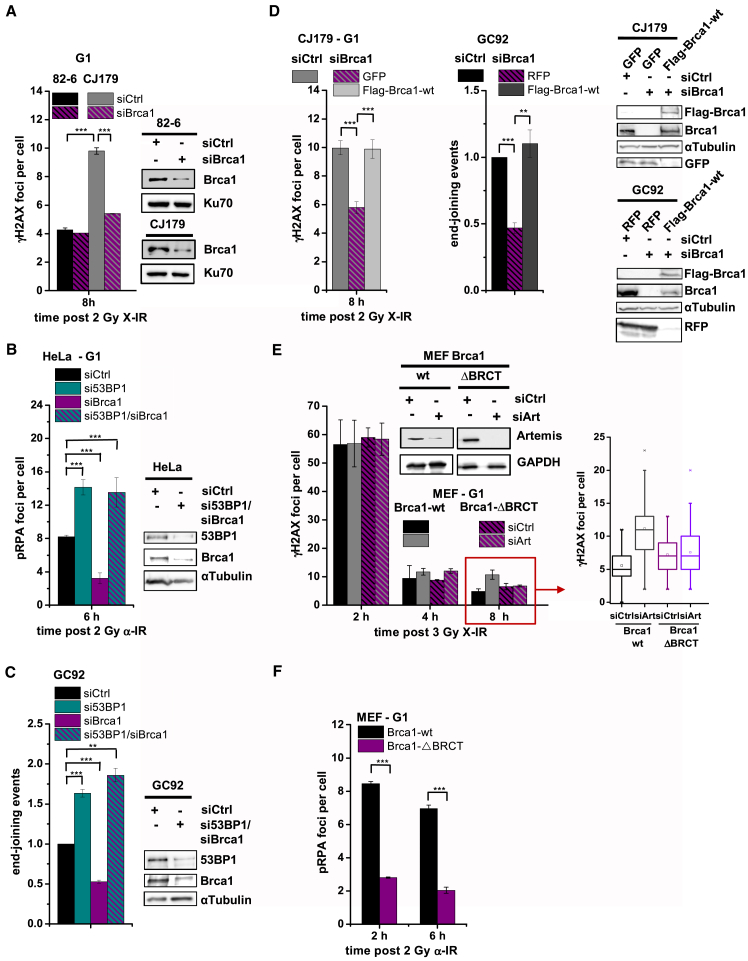
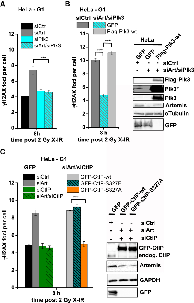
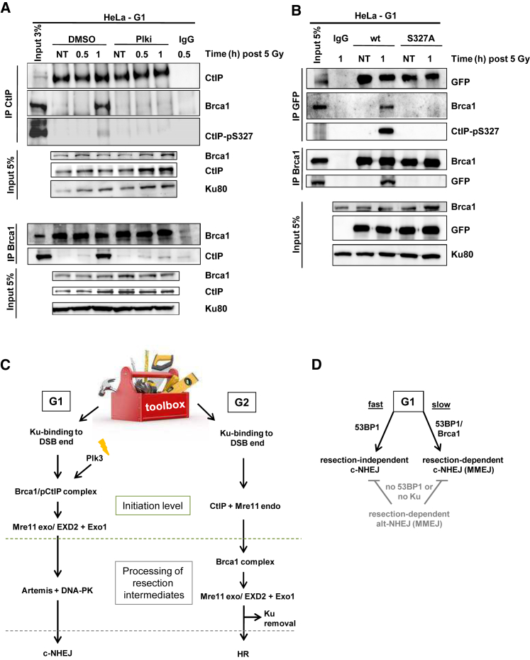
Canonical non-homologous end joining (c-NHEJ) repairs DNA double-strand breaks (DSBs) in G1 cells with biphasic kinetics. We show that DSBs repaired with slow kinetics, including those localizing to heterochromatic regions or harboring additional lesions at the DSB site, undergo resection prior to repair by c-NHEJ and not alt-NHEJ. Resection-dependent c-NHEJ represents an inducible process during which Plk3 phosphorylates CtIP, mediating its interaction with Brca1 and promoting the initiation of resection. Mre11 exonuclease, EXD2, and Exo1 execute resection, and Artemis endonuclease functions to complete the process. If resection does not commence, then repair can ensue by c-NHEJ, but when executed, Artemis is essential to complete resection-dependent c-NHEJ. Additionally, Mre11 endonuclease activity is dispensable for resection in G1. Thus, resection in G1 differs from the process in G2 that leads to homologous recombination. Resection-dependent c-NHEJ significantly contributes to the formation of deletions and translocations in G1, which represent important initiating events in carcinogenesis.

Keywords: DNA double-strand breaks; non-homologous end joining; nucleases; resection.


Figures

None
Graphical abstract
Figure 1
Figure 1
A Slow Artemis-Dependent c-NHEJ Process Promotes Translocation Formation in G1 (A) Left: FISH-stained G1 PCC spread from 82-6 control cells (dashed circle) fused with mitotic HeLa cells. Right: chromosome breaks and translocations in G1 PCC spreads. (B and C) Chromosome breaks (B) and translocations (C) in G1 82-6 cells treated with siLig1/3 or PARP inhibitor (PARPi). Data are mean ± SEM. (D) Chromosome breaks and translocations in G1 82-6 cells treated with DNA-PK inhibitor (DNA-PKi) 6 hr after X-IR. Data are mean ± SEM. (E) Chromosome breaks and translocations in G1 82-6 and Artemis-deficient CJ179 cells. Data are mean ± SEM. (F) γH2AX foci in G1 82-6 cells treated with DNA-PKi at various times after X-IR. Data are mean ± SEM. (G) γH2AX foci in G1 82-6, XLF-deficient 2BN and Lig4-mutated 411Br cells treated with PARPi. Data are mean ± SEM. (H) γH2AX foci in G1 82-6 and CJ179 cells treated with DNA-PKi 4 hr after X-IR. Data are mean ± SEM. See also Figures S1 and S2.
Figure 2
Figure 2
Artemis and CtIP Function during Slow DSB Repair and Promote Resection in G1 (A) γH2AX foci in G1 82-6 and CJ179 cells treated with siCtIP. Data are mean ± SEM. (B) γH2AX foci in CJ179 cells treated with siCtIP. Cells were transfected with GFP or GFP-CtIP-WT constructs, and γH2AX foci were analyzed in GFP+ G1 cells. Data are mean ± SEM. (C) Chromosome breaks in G1 82-6 and CJ179 cells treated with siCtIP. Data are mean ± SEM. (D) pRPA foci in G1 HeLa cells treated with siArtemis. Cells were transfected with GFP or cMyc-Artemis plasmids, and foci were analyzed in GFP/cMyc+ G1 cells. Data are mean ± SEM. (E) pRPA foci in G1 GC92 WT and CRISPR/Cas9-generated Artemis KO cells. Cells were transfected with GFP or cMyc-Artemis constructs, and pRPA foci were analyzed in GFP/cMyc+ G1 cells. Data are mean ± SEM. (F) pRPA foci in G1 GC92 WT and Artemis KO cells treated with siCtIP. Data are mean ± SEM. See also Figure S3.
Figure 3
Figure 3
Molecular Characterization of G1 Resection (A) Schematic of the NHEJ reporter assay. The repair of two I-SceI-induced DSBs can result in loss of the intervening fragment, which is detected by a CD4+ signal (Rass et al., 2009). CD4+ clones were amplified by PCR (green arrows) across the repair site and sequenced. Repair of the two DSBs can also occur without loss of the intervening fragment, which escapes detection. (B) γH2AX foci in GC92 WT and Artemis KO cells treated with siDNA-PKcs or siCtIP. Cells were transfected with I-SceI, and foci were scored in I-SceI+ and I-SceI cells (identified by immunofluorescence [IF] against I-SceI). Data are mean ± SEM. (C) End joining events in GC92 WT and Artemis KO cells containing the NHEJ reporter substrate. Cells were transfected with RFP or cMyc-Artemis constructs. Events were quantified by the fraction of CD4+ and RFP/cMyc+cells relative to all RFP/cMyc+cells, and results were normalized to WT cells. Data are mean ± SEM. (D) End joining events in GC92 WT and Artemis KO cells treated with siCtIP. Data are mean ± SEM. (E) Distribution of deletion sizes obtained from the sequence analysis of GC92 WT and siLig1/3-treated cells. nt, nucleotide. (F) End joining events in GC92 cells treated with siKu70/80, siLig4, siLig1/3, or siDNA-PKcs. Data are mean ± SEM. See also Table S1.
Figure 4
Figure 4
Similar and Distinct Nuclease Requirements for Resection in G1 versus G2 (A–C) γH2AX foci in G1 82-6 and CJ179 cells (A), pRPA foci in G1 HeLa cells (B), and end-joining events in GC92 cells (C). Cells were treated with an Mre11 endo- or exonuclease inhibitor, siEXD2, siExo1, or siBLM/siDNA2. Data are mean ± SEM. (D) γH2AX foci in G1 CJ179 and end joining events in GC92 cells. Cells were treated with siExo1 and transfected with GFP, RFP, or FLAG-Exo1-WT constructs, and GFP+, RFP+ or FLAG+ cells were analyzed. Data are mean ± SEM. (E) Model for DSB end resection in G1 and G2. DNA-PKcs binding to Ku was omitted for clarity. See also Figure S4.
Figure 5
Figure 5
Brca1 and 53BP1 Together Promote Resection-Dependent Slow DSB Repair in G1 (A) γH2AX foci in G1 82-6 and CJ179 cells treated with siBrca1. Data are mean ± SEM. (B) pRPA foci in G1 HeLa cells treated with si53BP1 and/or siBrca1. Data are mean ± SEM. (C) End joining events in GC92 cells treated with si53BP1 and/or siBrca1. Data are mean ± SEM. (D) γH2AX foci in G1 CJ179 and end joining events in GC92 cells. Cells were treated with siBrca1 and transfected with GFP, RFP or FLAG-Brca1-WT constructs, and GFP+, RFP+ or FLAG+ cells were analyzed. Data are mean ± SEM. (E) γH2AX foci in G1 Brca1-WT and Brca1-ΔBRCT MEFs treated with siArtemis. Data are mean ± SEM. (F) pRPA foci in G1 Brca1-WT and Brca1-ΔBRCT MEFs. Data are mean ± SEM. See also Figure S5.
Figure 6
Figure 6
Plk3 Is Required for Resection-Dependent Slow DSB Repair in G1 (A) γH2AX foci in G1 HeLa cells treated with siArtemis and/or siPlk3. Data are mean ± SEM. (B) γH2AX foci in G1 HeLa cells treated with siArtemis/siPlk3 and transfected with GFP or FLAG-Plk3-WT constructs, and GFP+ or FLAG+ G1 cells were analyzed. Data are mean ± SEM. (C) γH2AX foci in G1 HeLa cells treated with siArtemis and/or siCtIP. Cells were transfected with GFP or GFP-CtIP constructs, and GFP+ G1-phase cells were analyzed. Data are mean ± SEM. See also Figure S6.
Figure 7
Figure 7
CtIP Phosphorylation at Ser327 by Plk3 Mediates Interaction with Brca1 in G1 (A) Interaction of CtIP and Brca1 in synchronized G1 HeLa cells treated with Plki. (B) Interaction of CtIP and Brca1 in G1 HeLa cells transfected with GFP-CtIP-WT or GFP-CtIP-S327A. Brca1 or GFP/CtIP was immunoprecipitated from cell extracts, and protein levels were analyzed. (C) Model summarizing the hierarchy of investigated factors involved in resection-dependent c-NHEJ in G1 in comparison with HR in G2. (D) Model for DSB repair pathway choice in G1 human cells. See also Figure S7.

References

    1. Barton O., Naumann S.C., Diemer-Biehs R., Künzel J., Steinlage M., Conrad S., Makharashvili N., Wang J., Feng L., Lopez B.S. Polo-like kinase 3 regulates CtIP during DNA double-strand break repair in G1. J. Cell Biol. 2014;206:877–894. - PMC - PubMed
    1. Beucher A., Birraux J., Tchouandong L., Barton O., Shibata A., Conrad S., Goodarzi A.A., Krempler A., Jeggo P.A., Löbrich M. ATM and Artemis promote homologous recombination of radiation-induced DNA double-strand breaks in G2. EMBO J. 2009;28:3413–3427. - PMC - PubMed
    1. Broderick R., Nieminuszczy J., Baddock H.T., Deshpande R.A., Gileadi O., Paull T.T., McHugh P.J., Niedzwiedz W. EXD2 promotes homologous recombination by facilitating DNA end resection. Nat. Cell Biol. 2016;18:271–280. - PMC - PubMed
    1. Chang H.H., Watanabe G., Lieber M.R. Unifying the DNA end-processing roles of the artemis nuclease: Ku-dependent artemis resection at blunt DNA ends. J. Biol. Chem. 2015;290:24036–24050. - PMC - PubMed
    1. Chanut P., Britton S., Coates J., Jackson S.P., Calsou P. Coordinated nuclease activities counteract Ku at single-ended DNA double-strand breaks. Nat. Commun. 2016;7:12889. - PMC - PubMed

Publication types

MeSH terms

Substances