Review
. 2017 May:173:19-33.
doi: 10.1016/j.pharmthera.2017.01.004. Epub 2017 Jan 27.

Cellular retinoid binding-proteins, CRBP, CRABP, FABP5: Effects on retinoid metabolism, function and related diseases

Affiliations
Review

Cellular retinoid binding-proteins, CRBP, CRABP, FABP5: Effects on retinoid metabolism, function and related diseases

Joseph L Napoli. Pharmacol Ther. 2017 May.

Abstract

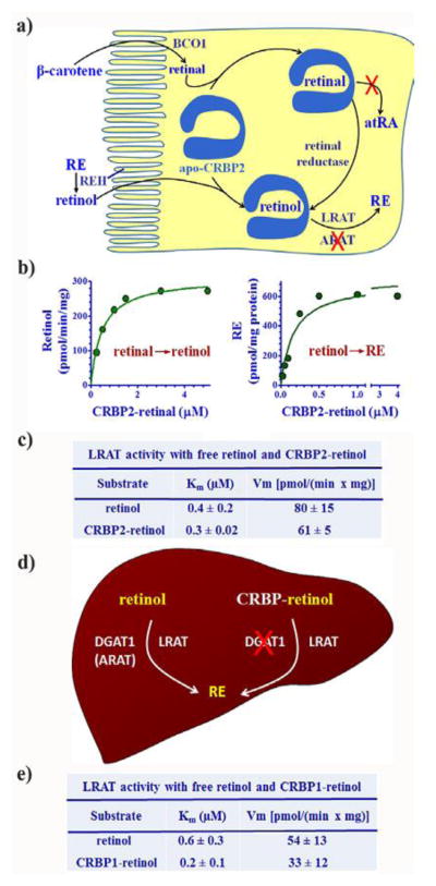
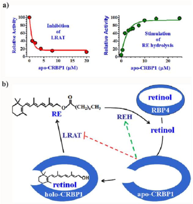
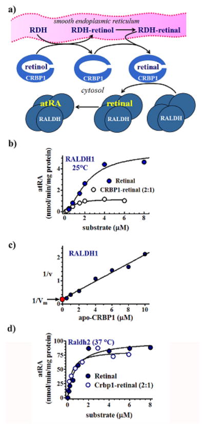
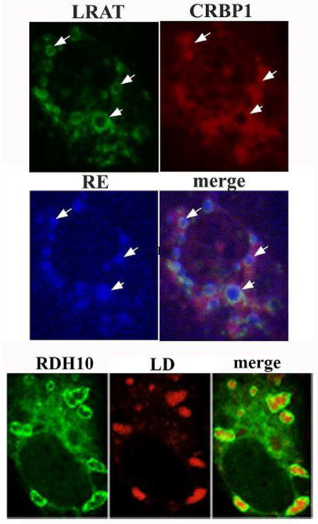
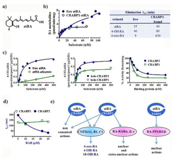
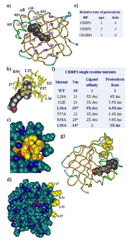
Cellular binding-proteins (BP), including CRBP1, CRBP2, CRABP1, CRABP2, and FABP5, shepherd the poorly aqueous soluble retinoids during uptake, metabolism and function. Holo-BP promote efficient use of retinol, a scarce but essential nutrient throughout evolution, by sheltering it and its major metabolite all-trans-retinoic acid from adventitious interactions with the cellular milieu, and by imposing specificity of delivery to enzymes, nuclear receptors and other partners. Apo-BP reflect cellular retinoid status and modify activities of retinoid metabolon enzymes, or exert non-canonical actions. High ligand binding affinities and the nature of ligand sequestration necessitate external factors to prompt retinoid release from holo-BP. One or more of cross-linking, kinetics, and colocalization have identified these factors as RDH, RALDH, CYP26, LRAT, RAR and PPARβ/δ. Michaelis-Menten and other kinetic approaches verify that BP channel retinoids to select enzymes and receptors by protein-protein interactions. Function of the BP and enzymes that constitute the retinoid metabolon depends in part on retinoid exchanges unique to specific pairings. The complexity of these exchanges configure retinol metabolism to meet the diverse functions of all-trans-retinoic acid and its ability to foster contrary outcomes in different cell types, such as inducing apoptosis, differentiation or proliferation. Altered BP expression affects retinoid function, for example, by impairing pancreas development resulting in abnormal glucose and energy metabolism, promoting predisposition to breast cancer, and fostering more severe outcomes in prostate cancer, ovarian adenocarcinoma, and glioblastoma. Yet, the extent of BP interactions with retinoid metabolon enzymes and their impact on retinoid physiology remains incompletely understood.

Keywords: Cellular retinoic acid binding-protein; Cellular retinol binding-protein; Retinal dehydrogenase; Retinoic acid; Retinoids; Retinol; Retinol dehydrogenase.


Conflict of interest statement

Conflict of Interest

The author declares that there are no conflicts of interest.

Figures

Fig. 1
Fig. 1
Impact of CRBP1 and 2 on retinal reduction and RE biosynthesis. a) Model of CRBP2 actions in the intestinal enterocyte that direct retinoids into RE formation and arrests atRA biosynthesis. A brush border REH converts dietary vitamin A esters into retinol for absorption (Rigtrup & Ong, 1992; Rigtrup et al., 1994a; Rigtrup et al., 1994b). Alternatively, metabolism of carotenoids produces retinal. CRBP2 directs retinoids into RE formation and prevents atRA biosynthesis (Kakkad & Ong, 1988). b) Michaelis-Menten relationships of CRBP2-mediated retinal reduction and retinol esterification (Ong et al., 1987; MacDonald & Ong, 1988b). c) Kinetic constants demonstrating holo-CRBP2 channeling retinol through protein-protein interactions into RE biosynthesis. d) Model showing CRBP1 and 2 actions in liver directing retinol to LRAT for RE formation (Ong et al., 1988; MacDonald & Ong, 1988a; Yost et al., 1988; Randolph et al., 1991; Herr & Ong, 1992). e) Kinetic constants that illustrate ability of holo-CRBP1 to deliver retinol through protein-protein interaction to LRAT for RE biosynthesis in liver.
Fig. 2
Fig. 2
Apo-CRBP1 enables mobilization of RE. a) Kinetics of apo-CRBP1 inhibiting LRAT and stimulating endogenous RE hydrolysis (Herr & Ong, 1992; Boerman & Napoli, 1991). b) Model relating actions of apo-CRBP1 to cellular retinoid homeostasis. Apo-CRBP1 acts as a sink to draw retinol into cells from the serum BP, RBP4, and also signals cell retinoid status. In the absence of apo-CRBP1, CRBP1 directs retinol into RE formation, while maintaining atRA biosynthesis. As apo-CRBP1 increases, inhibition of LRAT preserves remaining holo-CRBP1 to support atRA biosynthesis.
Fig. 3
Fig. 3
Contribution of CRBP1 to atRA homeostasis. a) Crosslinking with holo-CRBP1 identified RDH as members of the short-chain dehydrogenase/reductase gene family (SDR) by radio-iodinating this CRBP1 target enzyme (Boerman & Napoli, 1995). b) Michaelis-Menten kinetics confirmed ability of holo-CRBP1 to deliver retinol to RDH. The lack of change in Km or Vm with differences in the ratio CRBP1/retinol further confirmed direct interaction between holo-CRBP1 and RDH (Posch et al., 1991). c) Illustration of CRBP1’s impact on the amount of “free” retinol, calculated using a kd value of 2 nM. d) Kinetics of the reductase DHRS4 with free retinal or CRBP1-retinal (Lei et al., 2003). e) Inhibition of the DHRS4 reaction by apo-CRBP1, illustrating the mechanism of a decreased Vm with CRBP1-retinal as substrate shown in d. The filled blue symbols show that partial sequestration of retinal does not lower the reaction rate, indicating that both free and bound retinal are available as substrate. The red symbol shows the impact of equimolar amounts of BP and retinal, indicating ability of apo-CRBP1 to inhibit DHRS4.
Fig. 4
Fig. 4
Interactions of RALDH1 and 2 with CRBP1. A) Model depicting ability of cytosolic RALDH to access retinal generated in microsomes by RDH. CRBP1 acts as intermediate delivering retinal to the tetrameric RALDH for conversion into atRA. b) Kinetics of retinal generation from free retinal or CRBP1-bound retinal by RALDH1 (Penzes et al., 1997). A 2/1 ratio of CRBP1/retinal ensured total binding of retinal. The Vm from this ratio was lower than from retinal because apo-CRBP1 inhibits RALDH1. c) A plot allowing calculation of the Vm in the presence of an exact 1/1 ratio of CRBP1/retinal (red point). This approach showed that the Vm with no apo-CRBP1 present was 91% of the value using free retinal, which could occur only with direct interaction between CRBP1 and RALDH1. d) Michaelis-Menten kinetics illustrating channeling between CRBP1 and RALDH2, and lack of inhibition of RALDH2 by apo-CRBP1 (Wang et al., 1996).
Fig. 5
Fig. 5
LRAT, RDH10 and CRBP1 co-localize around LD during RE biosynthesis. Confocal microscopy of LRAT, CRBP1 and RDH10 with fluorescent reporters (Jiang & Napoli, 2012; Jiang & Napoli, 2013). LRAT localizes around LD containing RE, as does CRBP1. White arrows designate LD. RDH10 also concentrates on LD surfaces. This places LRAT and RDH10 at a major source of substrate for their reactions and also places the enzymes with the substrate delivery vehicle CRBP1. CRBP1 does not associate as tightly with LD as the two enzymes, consistent with its function of conducting retinol from the cell membrane to LD. These data suggest that LD act as organelles of hormone generation.
Fig. 6
Fig. 6
Functions of atRA BP. a) Structure of atRA designating two of the carbons oxidized by CYP26, C4 and C18. b) Michaelis-Menten kinetics revealing the differences between catabolism of free atRA vs. CRABP1-bound atRA. The reaction with BP was done with a 3/1 ratio of total CRABP1 to atRA to ensure lack of free atRA, yet enhanced the reaction rate at low concentrations of substrate. The reaction with BP-bound atRA followed typical Michaelis-Menten kinetics, whereas that of unbound atRA seemed unsaturable at concentrations that exceed the concentrations of atRA in tissues. The table shows the effects of binding atRA and two of its metabolites on their rates of catabolism, suggesting high concentrations of catabolites bound to CRABP contribute to atRA toxicity by blocking further catabolism of atRA (Fiorella & Napoli, 1991, 1994). c) Rate of 4-OH-atRA biosynthesis from free atRA or albumin bound atRA showing that not all protein-bound atRA behaves similar to CRABP-bound atRA. Michaelis-Menten kinetics illustrating protein-protein interactions between recombinant CYP26B1 and CRABP1 and 2. Impact of CRABP1 and 2 on the ability of recombinant CYP26B1 to catalyze atRA catabolism, indicating inhibition of CRP26B1 by the two atRA BP (Nelson et al., 2016). d) Effect of increasing the RAR concentration on the rate of transfer of atRA from BP. The decrease in the transfer rate indicates that only CRABP2 directly interacts with RAR (Dong et al., 1999). e) Summary of CRABP and FABP5 functions.
Fig. 7
Fig. 7
Structure-activity relationships of retinoid BP. a) Ribbon diagram of CRBP1 illustrating the entrance portal, key residues, orientation of retinol in the binding pocket, and the structure of the β-clam (Cowan et al., 1993; Franzoni et al., 2002; Lu et al., 2003; Careri et al., 2006; Mittag et al., 2006; Franzoni et al., 2010; 2016 Silvaroli et al., 2016). Red shows the hydroxyl group of retinol. Structure PDB ID 5HBS, NCBI. b) Partial structure focusing on interactions of entrance portal residues with each other and with the β-ionone ring. Red depicts the retinol hydroxyl group. c) Partial space-filling structure illustrating close contacts of residues in the entrance portal. Red illustrates the β-ionone ring. d) Total space-filling structure of CRBP1 showing sequestration of retinol from the cellular milieu, and residues that project from the αII helix out from the BP, which serve as putative “handles” that allow enzymes to tease retinol from the BP. Red indicates the β-ionone ring. e) Differences in proteolysis rates between apo- and holo-BP. BP were exposed to the endopeptidase Arg-C (clostripain). Degrees of proteolysis were analyzed by SDS-PAGE: scale: 0, no proteolysis; 1, low (<50%); 2, moderate (>50%); 3, complete. These data were obtained after 1 hour incubation, but holo-forms remained protease resistant up to 20 hours (Jamison et al., 1994). f) Effects of point mutations on ability of CRBP1 to deliver retinol to RDH, ligand affinity and rigidity (proteolysis rate) (Penzes and Napoli, 1999). g) Structure of CRABP1, which folds similarly to CRABP2, showing the carboxyl group (red) of atRA and the location of the three residues that enable RAR binding in CRABP2. Structure PDB ID 1CBR, NCBI.

Similar articles

Cited by

References

    1. Akhtar S, Ahmed A, Randhawa MA, Atukorala S, Arlappa N, Ismail T, Ali Z. Prevalence of vitamin A deficiency in South Asia: causes, outcomes, and possible remedies. Journal of Health, Population, and Nutrition. 2013;31(4):413–423. - PMC - PubMed
    1. Allegretto EA, McClurg MR, Lazarchik SB, Clemm DL, Kerner SA, Elgort MG, … Heyman RA. Transactivation properties of retinoic acid and retinoid X receptors in mammalian cells and yeast. Correlation with hormone binding and effects of metabolism. The Journal of Biological Chemistry. 1993;268(35):26625–26633. - PubMed
    1. Allenby G, Bocquel MT, Saunders M, Kazmer S, Speck J, Rosenberger M, … Chambon P. Retinoic acid receptors and retinoid X receptors: interactions with endogenous retinoic acids. Proceedings of the National Academy of Sciences of the United States of America. 1993;90(1):30–34. - PMC - PubMed
    1. Arnold SE, Kent T, Hogarth CA, Schlatt S, Prasad B, Haenisch M, Walsh T, Muller CH, Griswold MD, Amory JK, Isoherranen N. Importance of ALDH1A enzymes in determining human testicular retinoic acid concentrations. The Journal of Lipid Research. 2015;56:342–357. - PMC - PubMed
    1. Barbus S, Tews B, Karra D, Hahn M, Radlwimmer B, Delhomme N, … Lichter P. Differential retinoic acid signaling in tumors of long- and short-term glioblastoma survivors. Journal of the National Cancer Institute. 2011;103(7):598–606. - PubMed

MeSH terms