. 2016:17:117-123.

Therapeutic potential of the heme oxygenase-1 inducer hemin against Ebola virus infection

Affiliations

Therapeutic potential of the heme oxygenase-1 inducer hemin against Ebola virus infection

Hanxia Huang et al. Curr Trends Immunol. 2016.

Abstract

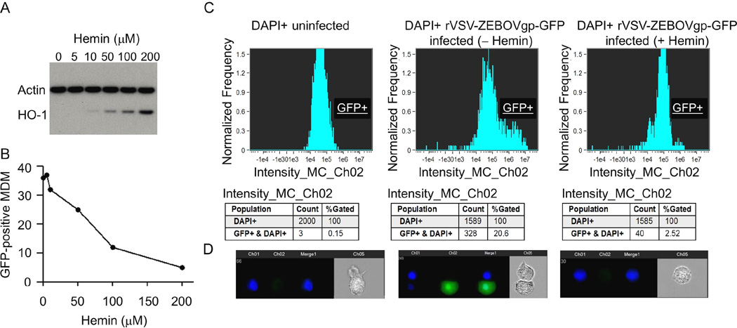
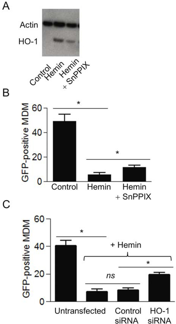
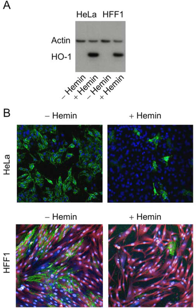
Promising drugs to treat Ebola virus (EBOV) infection are currently being developed, but so far none has shown efficacy in clinical trials. Drugs that can stimulate host innate defense responses may retard the progression of EBOV disease. We report here the dramatic effect of hemin, the natural inducer of the heme catabolic enzyme heme oxygenase-1 (HO-1), in the reduction of EBOV replication. Treatment of primary monocyte-derived macrophages (MDM), Vero E6 cells, HeLa cells, and human foreskin fibroblasts (HFF1) with hemin reduced EBOV infection by >90%, and showed minimal toxicity to infected cells. Inhibition of HO-1 enzymatic activity and silencing HO-1 expression prevented the hemin-mediated suppression of EBOV infection, suggesting an important role for induction of this intracellular mediator in restricting EBOV replication. The inverse correlation between hemin-induced HO-1 and EBOV replication provides a potentially useful therapeutic modality based on the stimulation of an innate cellular response against Ebola infection.

Keywords: Ebola; heme oxygenase-1; hemin; monocytes/macrophages.


Conflict of interest statement

STATEMENT The authors declare no conflict of interest.

Figures

Fig. 1
Fig. 1
Hemin treatment inhibits EBOV infection of monocyte-derived macrophages (MDM). (A) Western blot analysis of hemin-treated MDM. Cells were incubated with the indicated concentrations of hemin, cellular proteins were separated on a sodium dodecyl sulfate (SDS)-polyacrylamide gel, transferred to polyvinylidene difluoride (PVDF) nitrocellulose membrane, and probed simultaneously with HO-1 and actin antibodies. (B) Data demonstrating reduction of rVSV-EBOVgp-GFP replication in MDM treated with increased concentrations of hemin, expressed as the proportion of cells positive for GFP. (C) Quantitative analysis of rVSV-EBOVgp-GFP-infected MDM in the absence or presence of 100 µM hemin by ImageStream. (D) Representative cell images of DAPI detection and rVSV-EBOVgp-GFP-positive cells. A total of 10,000 cell events were collected from each of the samples for data analysis, and fluorescence intensity was determined by IDEAS v5.0 software (Amnis Corporation, Seattle, WA).
Fig. 2
Fig. 2
(A) Hemin-induced HO-1 protein expression in MDM remaining after treatment with the HO-1 enzyme inhibitor SnPPIX. Cells were cultured in the presence of 10 µM SnPPIX and 25 µM hemin for 24 hours and examined for the expression of HO-1 and actin by Western blot analysis using mouse-anti-human HO-1 monoclonal antibody and rabbit-anti-human actin polyclonal antibody. (B) Inhibition of HO-1 activity attenuates hemin-induced protection of MDM against rVSV-EBOVgp-GFP infection. MDM were pre-treated with 10 µM SnPPIX for 2 hours followed by treatment for 24 hours with 25 µM hemin. The cells were then infected with rVSV-EBOVgp-GFP, and after 24 hours, infected cells were quantified by fluorescence microscopy. (C) rVSV-EBOVgp-GFP replication in Vero E6 cells transfected with control siRNA and HO-1 siRNA. Untransfected and transfected cells were infected in the absence or presence of 100 µM hemin for 48 hours, and examined for infectivity by fluorescence microscopy as described in Materials and Methods. Data from two independent experiments are presented as mean ± SEM. *P<0.05; ns: not significant.
Fig. 3
Fig. 3
(A) Western blot analysis of hemin-treated HeLa and HFF1 cells. HO-1 expression in total-protein lysates isolated from HeLa cells and HFF1 cells cultured for 24 hours in the absence or presence of 100 µM hemin. Cells were incubated with 100 µM hemin, cellular proteins were separated on a SDS-polyacrylamide gel, transferred to PVDF nitrocellulose membrane, and probed simultaneously with HO-1 and actin antibodies. (B) Confocal microscopy of HeLa and HFF1 cells infected with EBOV under BSL-4 conditions in the absence or presence of 100 µM hemin. Images were acquired using the PE Opera confocal platform with a 10× objective. The data were analyzed using Acapella software. Green: Ebola infection; blue: nuclei (DRAQ5 stain); magenta: cytoplasm (HFF1 cells).
Fig. 4
Fig. 4
Dose response to hemin treatment of EBOV-infected HeLa and HFF1 cells. HeLa cells (panel A) and HFF1 cells (panel B) were infected with Kikwit-95 Zaire strain of Ebola virus under BSL-4 conditions for 48 hours in the presence of the indicated concentrations of hemin. It gave activity with EC50 = 17–27 µM in HeLa cells and CC50 > 100 µM and EC50 = 26–30 µM in HFF1 and CC50 > 100 µM, n = 4 done in two independent experiments. Cellular viability of the infected cells at various hemin concentrations are illustrated in panels C and D.

References

    1. Sanchez A, Geisbert TW, Feldmann H. Filoviridae: Marburg and Ebola Viruses. Philadelphia: Fields Virology Lippincott-Williams & Wilkins; 2007.
    1. Ascenzi P, Bocedi A, Heptonstall J, Capobianchi MR, Caro AD, Mastrangelo E, Bolognesi M, Ippolito G. Mol. Aspects Med. 2008;29:151–185. - PubMed
    1. Zhang Y, Feng S, Cowling BJ. Lancet. 2016;387:509. - PubMed
    1. Fang LQ, Yang Y, Jiang JF, Yao HW, Kargbo D, Li XL, Jiang BG, Kargbo B, Tong YG, Wang YW, Liu K, Kamara A, Dafae F, Kanu A, Jiang RR, Sun Y, Sun RX, Chen WJ, Ma MJ, Dean NE, Thomas H, Longini IM, Jr, Halloran ME, Cao WC. Proc. Natl. Acad. Sci. USA. 2016;113:4488–4493. - PMC - PubMed
    1. Moll R, Reece S, Cosford P, Kessel A. Br. Med. Bull. 2016;117:15–23. - PubMed

LinkOut - more resources