. 2017:2017:6834692.
doi: 10.1155/2017/6834692. Epub 2017 Jan 4.

An Assessment of Vitreous Degeneration in Eyes with Vitreomacular Traction and Macular Holes

Affiliations

An Assessment of Vitreous Degeneration in Eyes with Vitreomacular Traction and Macular Holes

Quraish Ghadiali et al. J Ophthalmol. 2017.

Abstract

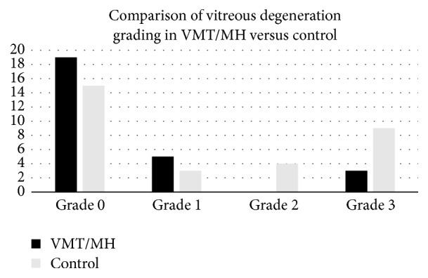
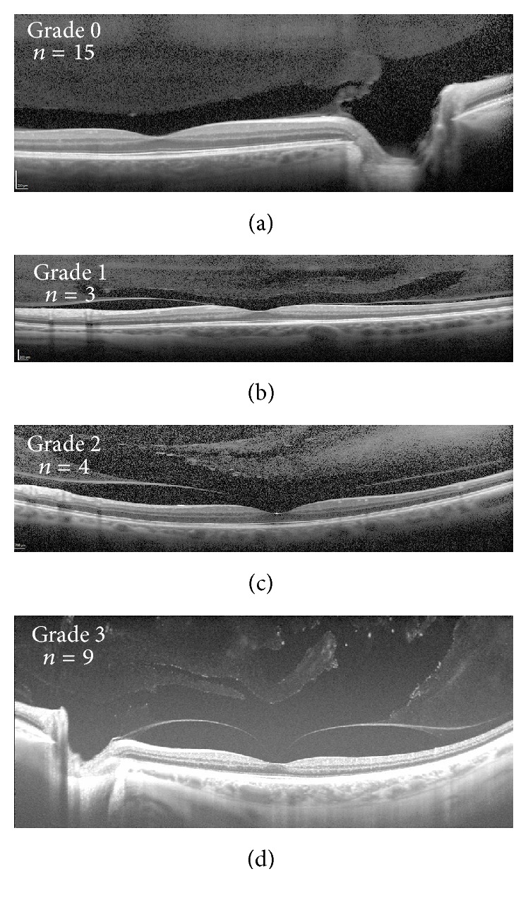
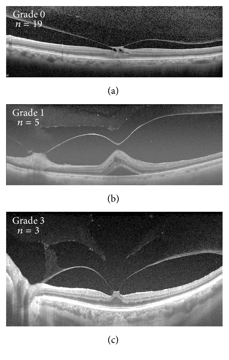
Purpose. To compare the stages of vitreous degeneration in patients with vitreomacular traction (VMT) and macular holes (MH). Methods. A retrospective study was performed analyzing stages of vitreous degeneration of eyes with VMT or MH using swept-source optical coherence tomography (SS-OCT) and spectral-domain optical coherence tomography (SD-OCT). An analogous review was performed on a control group of eyes with contralateral posterior vitreous detachments. Thirty-four eyes with VMT/MH and 39 control eyes were reviewed. Results. Twenty-seven VMT/MH eyes and 31 control eyes were included. Eyes with VMT/MH demonstrated significantly earlier stages of vitreous degeneration when compared to the control group (p = 0.048) despite significantly greater age (p = 0.032). Conclusions. Vitreoretinal interface disease is more often associated with a formed vitreous and an intact premacular bursa. This is contrary to previous assumptions implicating degeneration of vitreous as a precipitating factor of interface disease when in conjunction with abnormal vitreomacular separation.


Conflict of interest statement

No conflicting relationship exists for any author.

Figures

Figure 1
Figure 1
Distribution of vitreous grades in vitreomacular traction/macular hole (VMT/MH) eyes and control eyes. The VMT/MH eyes demonstrated significantly earlier degrees of vitreous degeneration when compared to the control group (p = 0.048).
Figure 2
Figure 2
Examples of vitreous grading in eyes from patients with a posterior vitreous detachment in the contralateral eye. Spectral-domain optical coherence tomography (a to c) and swept-source optical coherence tomography (d) examples of grade 0 (a), 1 (b), 2 (c), and 3 (d) vitreous degeneration. Number of eyes demonstrating each vitreous grade included in respective panels.
Figure 3
Figure 3
Examples of vitreous grading in eyes from patients with vitreomacular traction/macular hole (VMT/MH). Spectral-domain optical coherence tomography (a) and swept-source optical coherence tomography (b and c) examples of grade 0 (a), 1 (b), and 3 (c) vitreous degeneration. Number of eyes demonstrating each vitreous grade included in respective panels. Note that no patient in the VMT/MH group demonstrated grade 2 vitreous degeneration.
Figure 4
Figure 4
Vitreous degeneration force diagram: Shown is a schematic horizontal section of an eye with early stages of vitreous degeneration with formed vitreous (a). The forces are transmitted to the vitreoretinal interface by more central, formed vitreous without buffering from compartmentalization. Advanced vitreous degeneration with compartmentalization of the formed vitreous by liquid spaces may buffer the transmission of these forces (b).

References

    1. Sebag J. Anomalous posterior vitreous detachment: a unifying concept in vitreo-retinal disease. Graefe's Archive for Clinical and Experimental Ophthalmology. 2004;242(8):690–698. doi: 10.1007/s00417-004-0980-1. - DOI - PubMed
    1. Schaal K. B., Pang C. E., Pozzoni M. C., Engelbert M. The premacular bursa's shape revealed in vivo by swept-source optical coherence tomography. Ophthalmology. 2014;121(5):1020–1028. doi: 10.1016/j.ophtha.2013.11.030. - DOI - PubMed
    1. Itakura H., Kishi S., Li D., Akiyama H. Observation of posterior precortical vitreous pocket using swept-source optical coherence tomography. Investigative Ophthalmology and Visual Science. 2013;54(5):3102–3107. doi: 10.1167/iovs.13-11769. - DOI - PubMed
    1. Pang C. E., Schaal K. B., Engelbert M. Association of prevascular vitreous fissures and cisterns with vitreous degeneration as assessed by swept source optical coherence tomography. Retina. 2015;35(9):1875–1882. doi: 10.1097/IAE.0000000000000540. - DOI - PubMed
    1. Barteselli G., Bartsch D.-U., El-Emam S., et al. Combined depth imaging technique on spectral-domain optical coherence tomography. American Journal of Ophthalmology. 2013;155(4):727.e1–732.e1. doi: 10.1016/j.ajo.2012.10.019. - DOI - PMC - PubMed

LinkOut - more resources