. 2017:2017:3795950.
doi: 10.1155/2017/3795950. Epub 2017 Jan 4.

Hypothyroidism Reduces the Size of Ovarian Follicles and Promotes Hypertrophy of Periovarian Fat with Infiltration of Macrophages in Adult Rabbits

Affiliations

Hypothyroidism Reduces the Size of Ovarian Follicles and Promotes Hypertrophy of Periovarian Fat with Infiltration of Macrophages in Adult Rabbits

J Rodríguez-Castelán et al. Biomed Res Int. 2017.

Abstract

Ovarian failure is related to dyslipidemias and inflammation, as well as to hypertrophy and dysfunction of the visceral adipose tissue (VAT). Although hypothyroidism has been associated with obesity, dyslipidemias, and inflammation in humans and animals, its influence on the characteristics of ovarian follicles in adulthood is scarcely known. Control and hypothyroid rabbits were used to analyze the ovarian follicles, expression of aromatase in the ovary, serum concentration of lipids, leptin, and uric acid, size of adipocytes, and infiltration of macrophages in the periovarian VAT. Hypothyroidism did not affect the percentage of functional or atretic follicles. However, it reduced the size of primary, secondary, and tertiary follicles considered as large and the expression of aromatase in the ovary. This effect was associated with high serum concentrations of total cholesterol and low-density lipoprotein cholesterol (LDL-C). In addition, hypothyroidism induced hypertrophy of adipocytes and a major infiltration of CD68+ macrophages into the periovarian VAT. Our results suggest that the reduced size of ovarian follicles promoted by hypothyroidism could be associated with dyslipidemias, hypertrophy, and inflammation of the periovarian VAT. Present findings may be useful to understand the influence of hypothyroidism in the ovary function in adulthood.


Conflict of interest statement

Authors disclose that there are no financial or personal relationships with other people or organizations that could inappropriately bias or influence this work.

Figures
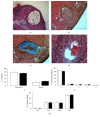
Figure 1
Figure 1
Follicle morphometry in control and hypothyroid rabbits. Atretic follicles: (a) cystic (Cyst), (b) invasive (Inv), (c) obliterative (Obli), and (d) residual (Res). Bars: (a) 500 μm, (b-c) 200 μm, and (d) 20 μm. Granulose cells, gc; theca cells, tc; and connective tissue, ct. (e) Percentage of ovarian functional and atretic follicles of control (open bars; n = 6) and hypothyroid (solid bars; n = 6) females rabbits. (f) Functional follicles were classified as primordial (P), primary (1), secondary (2), tertiary (3), and Graafian (G). (g) For atretic follicles, the different type of atresia mentioned above was analyzed. Data are mean ± SEM.
Figure 2
Figure 2
Diameter of primordial (a), primary (b), secondary (c), and tertiary (d) ovarian follicles of control (open bars; n = 6) and hypothyroid (solid bars; n = 6) females rabbits. The mean diameter for all (overall), large, and small follicles was analyzed. Large follicles were considered: primordial > 30 μm, primary > 50 μm, secondary > 100 μm, and tertiary > 250 μm. Data are mean ± SEM. P ≤ 0.05.
Figure 3
Figure 3
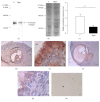
Expression of the cytochrome P450 aromatase in the ovary of control (C; open bar; n = 6) and hypothyroid (Hypo; solid bar; n = 6) rabbits. Representative immunoblot showing the expression of aromatase (a) and Ponceau's Red stained membrane (b). Relative expression of aromatase in C and Hypo groups (c). Data are mean ± SEM. ∗∗P < 0.01. Immunoreactivity of antiaromatase (+) was identified in granulosa cells of antral follicles (tertiary, tf; Graafian, gf) of control (d, e) and hypothyroid (f, g) ovaries. Nonlabeling was observed when the primary antibody was omitted (negative control; h). Bars: (d) 200 μm; (f) and (h) 100 μm; and (e) and (g) 20 μm. Granulose cells, gc; theca cells, tc.
Figure 4
Figure 4
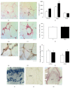
Histological characteristics of periovarian adipocytes of control (a and d) and hypothyroid (b and e) females rabbits. (c) The cross-sectional area of adipocytes (CSA; control in open bars; n = 6; hypothyroid in solid bars; n = 6). The average CSA for all (overall), large, and small adipocytes was analyzed. Large adipocytes were considered ≥4000 μm2. The presence of immune cells (polymorphonuclear cells; PMNC) around adipocytes was quantified (f). Data are mean ± SEM. P ≤ 0.05. Immunoreactivity of macrophages to CD163 and CD68 markers was analyzed (g, h, and l). The epithelium (ep) of the pancreatic duct in rabbits was considered as a negative control for anti-CD68 (i). The lack of the primary antibody (j) and the immunohistochemistry antiperilipin A (white arrows per+) in periovarian adipocytes were considered as negative controls for the secondary antibodies anti-mouse and anti-goat, respectively. Bars: (a), (b), (d), (e), and (j) = 50 μm; (g), (h), (i), and (k) = 20 μm.

Similar articles

Cited by

References

    1. Luque-Ramírez M., Álvarez-Blasco F., Uriol Rivera M. G., Escobar-Morreale H. F. Serum uric acid concentration as non-classic cardiovascular risk factor in women with polycystic ovary syndrome: effect of treatment with ethinyl-estradiol plus cyproterone acetate versus metformin. Human Reproduction. 2008;23(7):1594–1601. doi: 10.1093/humrep/den095. - DOI - PubMed
    1. Aytan A. N., Bastu E., Demiral I., Bulut H., Dogan M., Buyru F. Relationship between hyperandrogenism, obesity, inflammation and polycystic ovary syndrome. Gynecological Endocrinology. 2016;32(9):709–713. doi: 10.3109/09513590.2016.1155208. - DOI - PubMed
    1. Mannerås-Holm L., Leonhardt H., Kullberg J., et al. Adipose tissue has aberrant morphology and function in PCOS: enlarged adipocytes and low serum adiponectin, but not circulating sex steroids, are strongly associated with insulin resistance. Journal of Clinical Endocrinology and Metabolism. 2011;96(2):E304–E311. doi: 10.1210/jc.2010-1290. - DOI - PubMed
    1. Villa J., Pratley R. E. Adipose tissue dysfunction in polycystic ovary syndrome. Current Diabetes Reports. 2011;11(3):179–184. doi: 10.1007/s11892-011-0189-8. - DOI - PubMed
    1. Huang Z. H., Manickam B., Ryvkin V., et al. PCOS is associated with increased CD11c expression and crown-like structures in adipose tissue and increased central abdominal fat depots independent of obesity. The Journal of Clinical Endocrinology and Metabolism. 2013;98(1):E17–E24. doi: 10.1210/jc.2012-2697. - DOI - PMC - PubMed

LinkOut - more resources