. 2016;1(1):81-98.
doi: 10.20455/ros.2016.815.

Mitochondrial Electron Transport Chain-Derived Superoxide Exits Macrophages: Implications for Mononuclear Cell-Mediated Pathophysiological Processes

Affiliations

Mitochondrial Electron Transport Chain-Derived Superoxide Exits Macrophages: Implications for Mononuclear Cell-Mediated Pathophysiological Processes

Yunbo Li et al. React Oxyg Species (Apex). 2016.

Abstract

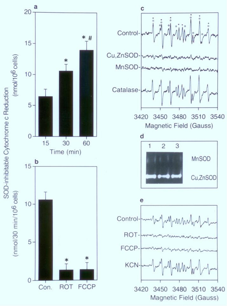
The involvement of mitochondrial electron transport chain (METC)-derived superoxide anion radical in cell protooncogene activation, mitogenic responses, and cancerous growth has recently received much attention. In order for METC-derived superoxide to participate in any of the above processes, its exit from mitochondria would be a critical step. Detection of intracellular superoxide showed that mitochondrial respiration is the major source of cellular superoxide in unstimulated or resting monocytes/macrophages. However, direct evidence for the exit of superoxide from mitochondria is presently lacking. Here we show that METC-derived superoxide does exit from mitochondria in unstimulated monocytes/macrophages. Release of superoxide was first found to occur with substrate-supported mitochondria isolated from these cells. We also observed the presence of extracellular superoxide with the intact unstimulated/resting cells. Extracellular superoxide was markedly diminished (>90%) by the mitochondrial inhibitor, rotenone, or the uncoupler, carbonylcyanide p-(trifluromethy) phenylhydrazone. Furthermore, cells with a deficient METC exhibited significant reduction (>90%) in extracellular superoxide, demonstrating that with intact cells METC-derived superoxide not only exits from mitochondria, but can be released extracellularly. Superoxide anion radical released from mitochondria could react with exogenous nitric oxide, forming peroxynitrite. Mitochondria-derived extracellular superoxide could also oxidize low-density lipoprotein (LDL). These results thus resolve any uncertainty on the ability of superoxide to exit from mitochondria. This study for the first time also identifies mitochondria as the major source of extracellular superoxide in unstimulated resting monocytes/macrophages, which has implications for the involvement of these mononuclear cells in various pathophysiological situations.

Keywords: Chemiluminescence; Electron paramagnetic resonance; Low-density lipoprotein; Macrophages; Mitochondrial electron transport chain; Monocytes; Mononuclear cells; Peroxynitrite; Reactive oxygen species; Superoxide.


Figures

FIGURE 1
FIGURE 1. Mitochondria as the major source of intracellular superoxide in unstimulated monocytes/macrophages
In panel a, a representative profile of lucigenin-derived CL elicited following incubation of unstimulated monocytes/macrophages with 1 µM lucigenin is shown. In panel b, intracellular superoxide production was detected by lucigenin-derived CL following incubation of unstimulated monocytes/macrophages with 1 µM lucigenin in the presence or absence of rotenone (ROT) (10 µM), FCCP (1 µM), or KCN (0.2 mM). Values in panel b represent the mean ± S.E. from 5 experiments.
Figure 2
Figure 2. Release of superoxide from isolated mitochondria
In panel a, intramitochondrial superoxide was detected by lucigenin-derived CL following incubation of succinate-supported mitochondria with 5 µM lucigenin in the presence or absence of MnTMPyP (5 µM) or Cu,ZnSOD (500 units/ml). Values represent the mean ± S.E. from 3 experiments. In panel b, formation of the DEPMPO-OH (o) was measured by EPR spectrometry following incubation of succinate-supported mitochondria with 10 mM DEPMPO in the presence or absence of Cu,ZnSOD (500 units/ml).
FIGURE 3
FIGURE 3. Presence of extracellular superoxide with unstimulated monocytes/macrophages and its diminution in the presence of mitochondrial inhibitors
In panel a, extracellular superoxide was measured by SOD-inhibitable cytochrome c reduction following incubation of unstimulated monocytes/macrophages for the indicated times. In panel b, extracellular superoxide was measured by SOD-inhibitable cytochrome c reduction following incubation of unstimulated monocytes/macrophages with rotenone (ROT) (10 µM) or FCCP (1 µM) for 30 min. In panel c, formation of DEPMPO-OOH (o) and DEPMPO-OH (*) was measured by EPR spectrometry following incubation of unstimulated monocytes/macrophages with 10 mM DEPMPO in the presence or absence of Cu,ZnSOD (500 units/ml), MnSOD (500 units/ml), or catalase (500 units/ml). In panel d, intracellular SOD activity was detected by SOD gel assay after incubation of unstimulated monocytes/macrophages with 500 units/ml Cu,ZnSOD (lane 2) or 500 units/ml MnSOD (lane 3) for 30 min. Lane 1 represents control cells. In panel e, formation of DEPMPO-spin adducts was detected by EPR spectrometry following incubation of unstimulated monocytes/macrophages with rotenone (ROT) (10 µM), FCCP (1 µM), or KCN (0.2 mM). In panels a and b, values represent the mean ± S.E. from 3 separate experiments.
FIGURE 4
FIGURE 4. Normal cell surface marker expression, intact plasma membrane NAD(P)H oxidase, and decreased mitochondrial respiration in monocytes/macrophages with a deficient METC
In panel a, substrate-supported mitochondrial oxygen consumption was measured polarographically in digitonin-permeabilized control cells and EB- or CAP-treated cells. Curves represent the average of three experiments. P/M, pyruvate/malate (6 mM/6 mM); ROT, rotenone (0.1 µM); Succ, succinate (6 mM); AA, antimycin A (0.1 µM); Asc./TMPD, ascorbate/tetramethyl-1,4-phenylenediamine (1 mM/0.2 mM). The sequence of addition of the above substrates and inhibitors was identical for control, EB-, and CAP-treated cells. In panel b, the expression of monocyte/macrophage surface markers, CD11b. CD14, and CD16 was measured flow cytometrically in control cells and EB- or CAP-treated cells. Values represent mean ± S.E. from 3 experiments. In panel c, production of superoxide by plasma membrane NAD(P)H oxidase following activation by TPA was measured by SOD-inhibitable cytochrome c reduction in control cells and EB- or CAP-treated cells. Values represent mean ± S.E. from five experiments. In panel d, the expression of cytosolic p47phox and p67phox was determined by immunoblotting (representative experiment of three).
FIGURE 5
FIGURE 5. Diminished presence of extracellular superoxide in unstimulated monocytes/macrophages with a deficient METC
In panel a, intracellular superoxide production was detected by lucigenin-derived CL in unstimulated control cells and EB-or CAP-treated cells. In panels b and c, presence of extracellular superoxide was measured by the SOD-inhibitable cytochrome c reduction assay and the DEPMPO-spin trapping, respectively, in unstimulated control cells and EB- or CAP-treated cells. In panels a and b, values represent the mean ± S.E. from at least 3 experiments.
FIGURE 6
FIGURE 6. Formation of peroxynitrite from reaction of nitric oxide with mitochondria-derived superoxide in unstimulated monocytes/macrophages
In panel a, formation of peroxynitrite was detected by luminol-derived CL following incubation of unstimulated monocytes/macrophages with 10 µM luminol in the presence or absence of the indicated concentrations of S-nitrosoglutathione and bicarbonate. Values represent the mean ± S.E. from at least 5 experiments. In panel b, formation of peroxynitrite was detected by luminol (10 µM)-derived CL following incubation of 100 µM S-nitrosoglutathione and 15 mM bicarbonate with unstimulated monocytes/macrophages in the presence or absence of Cu,ZnSOD (500 units/ml), rotenone (ROT) (10 µM), or FCCP (1 µM), or with EB- or CAP-treated cells. Values in both panels represent the mean ± S.E. from at least 3 experiments.
FIGURE 7
FIGURE 7. Oxidation of LDL by unstimulated monocytes/macrophages
LDL oxidation was detected by the TBARS assay following incubation of LDL with the unstimulated monocytes/macrophages in the presence or absence of Cu,ZnSOD (500 units/ml), catalase (500 units/ml), rotenone (ROT) (10 µM), or FCCP (1 µM), or with EB- or CAP-treated cells. Values represent the mean ± S.E. from at least 3 experiments.
FIGURE 8
FIGURE 8. Schematic illustration of the potential relevance of the exit of mitochondria-derived superoxide in mononuclear cell-mediated pathophysiological processes
O2• −, superoxide; NO, nitrioxide; ONOO, peroxynitrite. It should be noted that superoxide that exits from mitochondria may also play a role in normal cellular signaling and physiology.

Similar articles

Cited by

References

    1. Galli SJ, Borregaard N, Wynn TA. Phenotypic and functional plasticity of cells of innate immunity: macrophages, mast cells and neutrophils. Nat Immunol. 2011;12(11):1035–1044. - PMC - PubMed
    1. Babior BM, Kipnes RS, Curnutte JT. Biological defense mechanisms. The production by leukocytes of superoxide, a potential bactericidal agent. J Clin Invest. 1973;52(3):741–744. - PMC - PubMed
    1. Miles PR, Lee P, Trush MA, Van Dyke K. Chemiluminescence associated with phagocytosis of foreign particles in rabbit alveolar macrophages. Life Sci. 1977;20(1):165–170. - PubMed
    1. Fang FC. Antimicrobial reactive oxygen and nitrogen species: concepts and controversies. Nat Rev Microbiol. 2004;2(10):820–832. - PubMed
    1. Hansson GK. Inflammation, atherosclerosis, and coronary artery disease. N Engl J Med. 2005;352(16):1685–1695. - PubMed