

doi: 10.5301/jsrd.5000214.
Epub 2016 Oct 18.
Gastrointestinal Manifestations of Systemic Sclerosis
Affiliations
- PMID: 28133631
- PMCID: PMC5267349
- DOI: 10.5301/jsrd.5000214
Item in Clipboard
Gastrointestinal Manifestations of Systemic Sclerosis
J Scleroderma Relat Disord.
2016.
Abstract
In patients with systemic sclerosis (SSc), gastrointestinal (GI) tract involvement is almost universal. Any segment of the GI tract from mouth to anus can be involved, and GI symptoms are a frequent cause of morbidity. In severe cases, GI tract involvement can progress to the point of malnutrition requiring parenteral nutrition. GI tract involvement in SSc contributes to disease-related mortality although mostly as a co-morbidity rather than direct cause of death. The review is intended to help address challenges in the assessment and treatment of GI tract involvement in SSc.
Keywords: gastrointestinal; motility; scleroderma; systemic sclerosis.
Figures
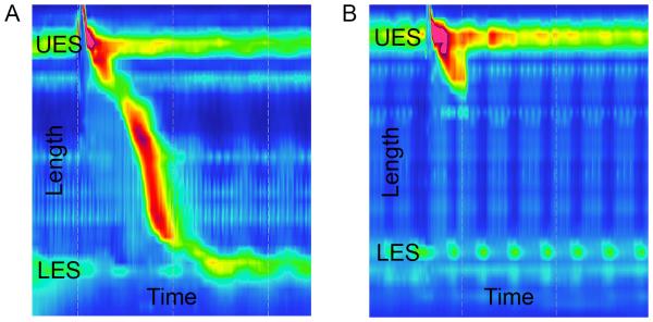
Esophageal manometry images for a normal control patient (A) and a patient with systemic sclerosis (B) are shown. The y-axis shows the length measured in the esophagus, including the location of the upper esophageal sphincter (UES) and lower esophageal sphincter (LES). The x-axis shows the time of the recording that captured one swallow. In (A), the diagonal region with topographical color change shows a normal swallow with distal propagation of esophageal peristalsis with time and corresponding LES relaxation. In (B), esophageal peristalsis is notably absent from the distal two-thirds of the esophagus, and resting LES pressure is low.
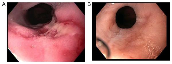
Endoscopic images of the distal esophagus are shown from the same patient with systemic sclerosis depicting grade C esophagitis (A) and healed esophagitis (B) after several weeks of proton-pump inhibitor therapy.
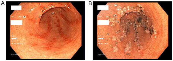
Endoscopic images from a patient with systemic sclerosis depicting gastric antral vascular ectasia before (A) and after (B) argon plasma coagulation therapy.
Similar articles
-
Severe gastrointestinal involvement in systemic sclerosis: report of five cases and review of the literature.Semin Arthritis Rheum. 2005 Feb;34(4):689-702. doi: 10.1016/j.semarthrit.2004.08.009. Semin Arthritis Rheum. 2005. PMID: 15692963 Review.
-
Management of gastrointestinal involvement in scleroderma.Curr Treatm Opt Rheumatol. 2015 Mar 1;1(1):82-105. doi: 10.1007/s40674-014-0005-0. Curr Treatm Opt Rheumatol. 2015. PMID: 26005632 Free PMC article.
-
Gastrointestinal manifestations of scleroderma: recent progress in evaluation, pathogenesis, and management.Curr Rheumatol Rep. 2012 Feb;14(1):22-9. doi: 10.1007/s11926-011-0217-3. Curr Rheumatol Rep. 2012. PMID: 22105546 Review.
-
Gastrointestinal involvement in systemic sclerosis: An updated review.Medicine (Baltimore). 2022 Nov 11;101(45):e31780. doi: 10.1097/MD.0000000000031780. Medicine (Baltimore). 2022. PMID: 36397401 Free PMC article. Review.
-
Gastrointestinal Manifestations, Malnutrition, and Role of Enteral and Parenteral Nutrition in Patients With Scleroderma.J Clin Gastroenterol. 2015 Aug;49(7):559-64. doi: 10.1097/MCG.0000000000000334. J Clin Gastroenterol. 2015. PMID: 25992813 Review.
Cited by
-
Effects of faecal microbiota transplantation on the small intestinal mucosa in systemic sclerosis.Rheumatology (Oxford). 2023 Aug 1;62(8):2918-2929. doi: 10.1093/rheumatology/kead014. Rheumatology (Oxford). 2023. PMID: 36688692 Free PMC article.
-
Expression of apoptotic and proliferation factors in gastric mucosa of patients with systemic sclerosis correlates with form of the disease.Sci Rep. 2019 Dec 5;9(1):18461. doi: 10.1038/s41598-019-54988-0. Sci Rep. 2019. PMID: 31804582 Free PMC article.
-
Gastrointestinal involvement in systemic sclerosis: Effects on morbidity and mortality and new therapeutic approaches.J Scleroderma Relat Disord. 2021 Feb;6(1):37-43. doi: 10.1177/2397198319891282. Epub 2019 Dec 20. J Scleroderma Relat Disord. 2021. PMID: 35382247 Free PMC article.
-
Fecal microbiome differs between patients with systemic sclerosis with and without small intestinal bacterial overgrowth.J Scleroderma Relat Disord. 2021 Oct;6(3):290-298. doi: 10.1177/23971983211032808. Epub 2021 Jul 24. J Scleroderma Relat Disord. 2021. PMID: 35382497 Free PMC article.
-
Evaluation of risk factors for pseudo-obstruction in systemic sclerosis.Semin Arthritis Rheum. 2019 Dec;49(3):405-410. doi: 10.1016/j.semarthrit.2019.05.005. Epub 2019 May 23. Semin Arthritis Rheum. 2019. PMID: 31202479 Free PMC article.
References
-
- Thoua NM, Bunce C, Brough G, Forbes A, Emmanuel AV, Denton CP. Assessment of gastrointestinal symptoms in patients with systemic sclerosis in a UK tertiary referral centre. Rheumatology (Oxford) 2010;49(9):1770–5. - PubMed
-
- Sandmeier B, Jager VK, Nagy G, Carreira PE, Tzankov A, Widuchowska M, et al. Autopsy versus clinical findings in patients with systemic sclerosis in a case series from patients of the EUSTAR database. Clin Exp Rheumatol. 2015;33(4 Suppl 91):S75–9. - PubMed
-
- Schmeiser T, Saar P, Jin D, Noethe M, Muller A, Soydan N, et al. Profile of gastrointestinal involvement in patients with systemic sclerosis. Rheumatol Int. 2012;32(8):2471–8. - PubMed
-
- Omair MA, Lee P. Effect of gastrointestinal manifestations on quality of life in 87 consecutive patients with systemic sclerosis. J Rheumatol. 2012;39(5):992–6. - PubMed
Grants and funding
LinkOut - more resources
Full Text Sources
Other Literature Sources