Review
. 2017 Jan;275(1):21-32.
doi: 10.1111/imr.12507.

The HIV-1 envelope glycoprotein structure: nailing down a moving target

Affiliations
Review

The HIV-1 envelope glycoprotein structure: nailing down a moving target

Andrew B Ward et al. Immunol Rev. 2017 Jan.

Abstract

Structure determination of the HIV-1 envelope glycoprotein (Env) presented a number of challenges, but several high-resolution structures have now become available. In 2013, cryo-EM and x-ray structures of soluble, cleaved SOSIP Env trimers from the clade A BG505 strain provided the first glimpses into the Env trimer fold as well as more the variable regions. A recent cryo-EM structure of a native full-length trimer without any stabilizing mutations had the same core structure, but revealed new insights and features. A more comprehensive and higher resolution understanding of the glycan shield has also emerged, enabling a more complete representation of the Env glycoprotein structure. Complexes of Env trimers with broadly neutralizing antibodies have surprisingly illustrated that most of the Env surface can be targeted in natural infection and that the neutralizing epitopes are almost all composed of both peptide and glycan components. These structures have also provided further evidence of the inherent plasticity of Env and how antibodies can exploit this flexibility by perturbing or even stabilizing the trimer to facilitate neutralization. These breakthroughs have stimulated further design and stabilization of Env trimers as well as other platforms to generate trimers that now span multiple subtypes. These Env trimers when used as immunogens, have led to the first vaccine-induced neutralizing antibodies for structural and functional analyses.

Keywords: HIV envelope structure; cryo-electron microscopy; epitope mapping; glycan shield; structure-based vaccine design; x-ray crystallography.


Conflict of interest statement

The authors declare no conflicts of interest.

Figures

Figure 1
Figure 1
The Env trimer architecture. (A) The 3 Å crystal structure of BG505 SOSIP Env (PDB 5CEZ). Different regions of the trimer described in the main text are colored accordingly: gp120 (gray), V1/V2 trimer apex (magenta), V3 loop (cyan), V4 loop (red), gp41 (brown), N‐terminal region of HR1 (HR1N) (green), and fusion peptide (yellow). Dashed lines indicate the locations of flexible, typically disordered regions in the trimer structures. The HR1N region (green) is also usually disordered in SOSIP structures, although fully resolved in 5CEZ. This same region in native Env trimers lacking the SOSIP mutations adopts a helical topology (PDB 5FUU). (B) Overlay of all Env trimer structures to date determined by cryo‐EM and x‐ray crystallography at <6 Å resolution (PDB: 3J5M, 4NCO, 4TVP, 4ZMJ, 5ACO, 5C7K, 5CEZ, 5CJX, 5D9Q, 5FUU, 5FYJ, 5FYK, 5FYL, 5I8H, 5JS9, 5JSA) demonstrates the conserved fold of the trimer, with small differences in the variable loops at the periphery of the trimer. The arrows indicate the fusion peptides from two different structures (5FUU, magenta and 5I8H, green) demonstrating its highly flexible nature
Figure 2
Figure 2
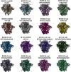
Overview of Env trimer structures solved to date at <6 Å resolution arranged in order of their structures being determined. First row: (PDB: 3J5M, 4NCO, 4TVP, 4ZMJ), Second row: (PDB: 5ACO, 5C7K, 5CEZ, 5CJX), Third row: (PDB: 5D9Q, 5FUU, 5FYJ, 5FYK), Fourth row: (PDB: 5FYL, 5I8H, 5JS9, 5JSA). The headers above the structures indicate the strain (clade A BG505, clade B JRFL, or clade G X1193.c1), the technique used to solve the structure (cryoEM or X‐ray), and the antibodies bound in the deposited structure (the Fabs are not shown for clarity). Most of the crystal structures have an antibody that targets the N332 supersite in gp120 (PGT122 or PGT128) and a gp41 or gp41/gp120 base‐binding antibody (35O22, 8ANC195, or VRC34), which also aid in crystallization. Antibodies can induce local structural changes within and proximal to their epitopes, particularly in the variable loops (V1‐V5), the fusion peptide, and glycans (data not shown). Some of these subtle differences are obvious in the images in the figure
Figure 3
Figure 3
Full‐length and soluble Env constructs. The almost complete native trimer with membrane‐proximal external region (MPER) and transmembrane domain (TMD) with only the C‐terminal domain deleted (ΔCT) is superimposed on a low resolution cryo‐ET reconstruction of Env on a viral membrane (EMD‐5019). The soluble cleaved BG505 trimer is shown on the bottom left with the SOSIP mutations. The NFL trimer construct is shown in the middle with a linker replacing the cleavage sequence between gp120 and gp41 with the I559P mutation form SOSIP and other trimer‐domain mutations added to expand the variety of strains and clades that can use this platform.84, 85 A further uncleaved UFO construct has a truncation and modification of HR1N as well as the SOS disulfide and a flexible linker region connecting gp120 and gp41 (bottom right)21
Figure 4
Figure 4
(A) Superposition of Env trimer structures determined to date with all resolved glycans colored green. While much of the surface of Env is covered by glycans, a few peptide epitopes are still accessible, most notably the CD4 binding site (dashed oval), although access is still highly restricted by the surrounding glycans. (B) The glycans resolved in the BG505 (PDB 2FYL) and JRFL (PBD 5FUU and 2FYK) Env trimers are displayed and colored according to the glycan site‐specific analyses of BG505 SOSIP Env in71 (high mannose: green, complex: magenta, high mannose and complex: orange, unknown: gray)

References

    1. Brand CM, Skehel JJ. Crystalline antigen from the influenza virus envelope. Nat New Biol. 1972;238:145–147. - PubMed
    1. Wiley DC, Skehel JJ. Crystallization and x‐ray diffraction studies on the haemagglutinin glycoprotein from the membrane of influenza virus. J Mol Biol. 1977;112:343–347. - PubMed
    1. Wrigley NG, Laver WG, Downie JC. Binding of antibodies to isolated haemagglutinin and neuraminidase molecules of influenza virus observed in the electron microscope. J Mol Biol. 1977;109:405–421. - PubMed
    1. Laver WG, Valentine RC. Morphology of the isolated hemagglutinin and neuraminidase subunits of influenza virus. Virology. 1969;38:105–119. - PubMed
    1. Wrigley NG, Skehel JJ, Charlwood PA, Brand CM. The size and shape of influenza virus neuraminidase. Virology. 1973;51:525–529. - PubMed

Publication types