. 2017 Mar;81(3):430-443.
doi: 10.1002/ana.24885. Epub 2017 Mar 20.

Typical and atypical pathology in primary progressive aphasia variants

Affiliations

Typical and atypical pathology in primary progressive aphasia variants

Edoardo G Spinelli et al. Ann Neurol. 2017 Mar.

Abstract

Objective: To characterize in vivo signatures of pathological diagnosis in a large cohort of patients with primary progressive aphasia (PPA) variants defined by current diagnostic classification.

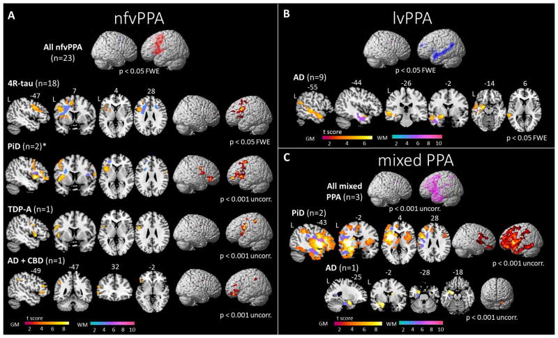
Methods: Extensive clinical, cognitive, neuroimaging, and neuropathological data were collected from 69 patients with sporadic PPA, divided into 29 semantic (svPPA), 25 nonfluent (nfvPPA), 11 logopenic (lvPPA), and 4 mixed PPA. Patterns of gray matter (GM) and white matter (WM) atrophy at presentation were assessed and tested as predictors of pathological diagnosis using support vector machine (SVM) algorithms.

Results: A clinical diagnosis of PPA was associated with frontotemporal lobar degeneration (FTLD) with transactive response DNA-binding protein (TDP) inclusions in 40.5%, FTLD-tau in 40.5%, and Alzheimer disease (AD) pathology in 19% of cases. Each variant was associated with 1 typical pathology; 24 of 29 (83%) svPPA showed FTLD-TDP type C, 22 of 25 (88%) nfvPPA showed FTLD-tau, and all 11 lvPPA had AD. Within FTLD-tau, 4R-tau pathology was commonly associated with nfvPPA, whereas Pick disease was observed in a minority of subjects across all variants except for lvPPA. Compared with pathologically typical cases, svPPA-tau showed significant extrapyramidal signs, greater executive impairment, and severe striatal and frontal GM and WM atrophy. nfvPPA-TDP patients lacked general motor symptoms or significant WM atrophy. Combining GM and WM volumes, SVM analysis showed 92.7% accuracy to distinguish FTLD-tau and FTLD-TDP pathologies across variants.

Interpretation: Each PPA clinical variant is associated with a typical and most frequent cognitive, neuroimaging, and neuropathological profile. Specific clinical and early anatomical features may suggest rare and atypical pathological diagnosis in vivo. Ann Neurol 2017;81:430-443.


Conflict of interest statement

Potential conflicts of interest

Nothing to report.

Figures

Figure 1
Figure 1
Flow chart showing inclusion/exclusion process of patients for the present study. Numbers of patients who underwent general cognitive, language and neuroimaging assessments are specified for each group. Abbreviations: AD= Alzheimer’s disease; GGT= globular glial tauopathy; lvPPA= logopenic variant PPA; MND= motor neuron disease; nfvPPA= non-fluent/agrammatic variant PPA; PiD= Pick’s disease; PSP= progressive supranuclear palsy; svPPA= semantic variant PPA; TDP-A, TDP-B, TDP-C= frontotemporal lobar degeneration with TDP-43 depositions type A, type B or type C.
Figure 2
Figure 2
Primary neuropathological diagnosis in primary progressive aphasia (PPA) clinical variants. Values are frequencies (percentages). Abbreviations: AD= Alzheimer’s disease; CBD= corticobasal degeneration; FTLD= frontotemporal lobar degeneration; GGT= globular glial tauopathy; lvPPA= logopenic variant PPA; nfvPPA= non-fluent/agrammatic variant PPA; PiD= Pick’s disease; PSP= progressive supranuclear palsy; svPPA= semantic variant PPA; tau 4R= FTLD-tau with 4 repeats.
Figure 3
Figure 3
Voxel-based morphometry analysis in patients with semantic variant of primary progressive aphasia (svPPA). Patterns of (A) cortical atrophy in all svPPA patients relative to healthy controls; (B) grey and white matter atrophy in each pathological subgroup relative to healthy controls; and (C) grey and white matter atrophy in svPPA patients with frontotemporal lobar degeneration due to tau (FTLD-tau) relative to those with TDP-43 depositions (FTLD-TDP) are shown, overlaid on sections or a three-dimensional rendering of a standard Montreal Neurologic Institute (MNI) brain. Slice labels refer to MNI coordinates. Abbreviations: FTLD= frontotemporal lobar degeneration; GGT= globular glial tauopathy; L= left; PiD= Pick’s disease; R= right; svPPA= semantic variant PPA; TDP-B= frontotemporal lobar degeneration with TDP-43 depositions type B; TDP-C= frontotemporal lobar degeneration with TDP-43 depositions type C.
Figure 4
Figure 4
Voxel-based morphometry analysis showing patterns of grey and white matter atrophy in patients with (A) non-fluent/agrammatic variant of primary progressive aphasia (nfvPPA), (B) logopenic variant of primary progressive aphasia (lvPPA) and (C) mixed PPA patients relative to healthy controls, according to pathological subgroups. All contrasts are overlaid on sections or a three-dimensional rendering of a standard Montreal Neurologic Institute (MNI) brain. Slice labels refer to MNI coordinates. *= a third nfvPPA patient with Pick’s disease showed selective right-sided atrophy (not displayed, see description in body text). Abbreviations: 4R-tau= frontotemporal lobar degeneration with 4-repeat tau depositions; AD= Alzheimer’s disease; FTLD= frontotemporal lobar degeneration; L= left; nfvPPA= non-fluent/agrammatic variant PPA; PiD= Pick’s disease; R= right; TDP-A= frontotemporal lobar degeneration with TDP-43 depositions type A.
Figure 5
Figure 5
Performance of support vector machine classification at a single-patient level is represented for patterns of grey matter (GM, first column), white matter (WM, second column) and a relative combination (GM+WM, third column). Colors and shapes indicate the pathological diagnosis (blue squares, PPA patients with FTLD-tau; green triangles, PPA with FTLD-TDP). Patients with “atypical” pathologies (i.e., svPPA-tau and nfvPPA-TDP) are filled in red. Patients are distributed along a vertical axis according to the probability of belonging to the FTLD-tau group (threshold >0.5) or the FTLD-TDP group (threshold <0.5). Abbreviations: FTLD= frontotemporal lobar degeneration; FTLD-tau= FTLD with tau depositions; FTLD-TDP= FTLD with TDP-43 depositions; nfvPPA= non-fluent/agrammatic variant PPA; svPPA= semantic variant PPA.

References

    1. Gorno-Tempini ML, Hillis AE, Weintraub S, et al. Classification of primary progressive aphasia and its variants. Neurology. 2011 Mar 15;76(11):1006–14. - PMC - PubMed
    1. Chare L, Hodges JR, Leyton CE, et al. New criteria for frontotemporal dementia syndromes: clinical and pathological diagnostic implications. Journal of neurology, neurosurgery, and psychiatry. 2014 Aug;85(8):865–70. - PubMed
    1. Mesulam M, Wicklund A, Johnson N, et al. Alzheimer and frontotemporal pathology in subsets of primary progressive aphasia. Annals of neurology. 2008 Jun;63(6):709–19. - PMC - PubMed
    1. Mesulam MM, Weintraub S, Rogalski EJ, Wieneke C, Geula C, Bigio EH. Asymmetry and heterogeneity of Alzheimer’s and frontotemporal pathology in primary progressive aphasia. Brain: a journal of neurology. 2014 Apr;137(Pt 4):1176–92. - PMC - PubMed
    1. Grossman M. Primary progressive aphasia: clinicopathological correlations. Nature reviews Neurology. 2010 Feb;6(2):88–97. - PMC - PubMed

MeSH terms